
A renal cell line with stable transporter expression
a renal cell line and transporter technology, applied in the field of drugdrug interaction and nephrotoxicity, can solve the problems of increasing the risk of ddis, reducing the number of patients, so as to achieve greater variation
- Summary
- Abstract
- Description
- Claims
- Application Information
AI Technical Summary
Benefits of technology
Problems solved by technology
Method used
Image

Examples
example 1
and Methods
[0105]Cell Culture
[0106]Conditionally immortalized proximal tubule epithelial cells (ciPTEC) were developed as described by Wilmer et al. with informed consent of the donors in accordance with the approved guidelines of the Radboud Institutional Review Board. (Wilmer et al., 2010) Cells were seeded 7 days prior to the experiment at their corresponding density (55,000 cells / cm2 for ciPTEC parent cells, 63,000 cells / cm2 for ciPTEC-OAT1 and 82,000 cells / cm2 for ciPTEC-OAT3) and grown for 1 day at 33° C. and 5% v / v CO2 to allow proliferation, enabled by the temperature-sensitive mutant of SV large T antigen (SV40T). Next, cells were cultured for 6 days at 37° C. and 5% v / v CO2 to stimulate differentiation and formation of an epithelial monolayer, described as ‘maturation’. Cells were cultured using Dulbecco's modified eagle medium (DMEM HAM's F12, Life Technologies, Paisly, UK), 5 μg / ml insulin, 5 μg / ml transferrin, 5 μg / ml selenium, 35 ng / ml hydrocortisone, 10 ng / ml epiderma...
example 2
al OAT Expression in ciPTEC
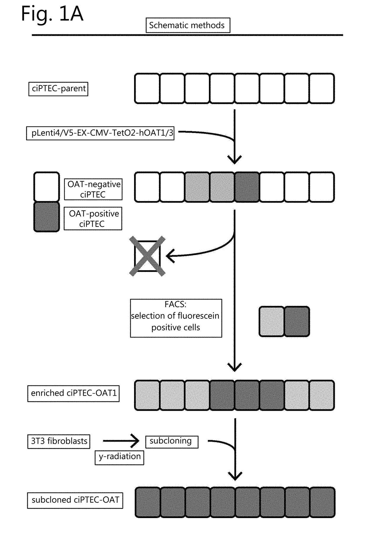
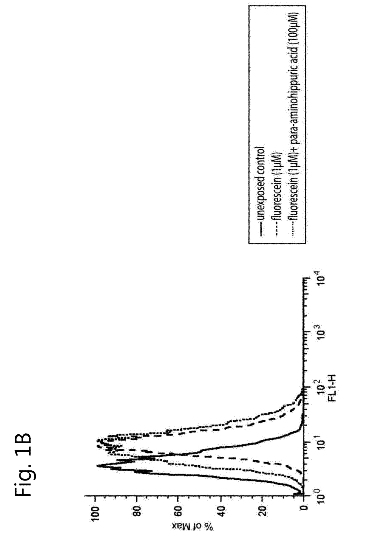
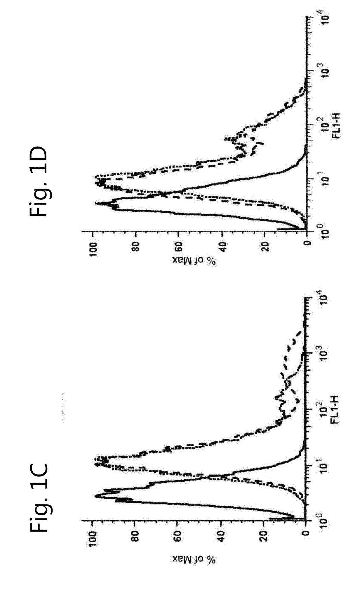
[0120]The absence of endogenous OAT1 and OAT3 expression in ciPTEC was demonstrated by exposure to fluorescein (1 μM) for 10 min, which did not increase the intracellular fluorescence intensity as measured by flow cytometry (FIG. 1B, dashed line). Therefore, OAT transporters were introduced separately by lentiviral transduction. A schematic overview of the experimental approach is provided in FIG. 1A. The transporter genes SLC22A6 and SLC22A8 were cloned under regulation of a CMV promoter and a TetO2 site to conditionally induce their expression. Remarkably, basal expression and function upon transduction of both OAT transporters was positive without tetracycline induction, and was not influenced by this inducer (data not shown). Fluorescein uptake capacity (without induction by tetracycline) was used to discriminate between successfully transduced cells and non-transduced cells, reflected by two sub-populations in the flow cytometer histograms (ciPTEC-OAT...
example 3
raction at the Site of OAT1 and OAT3
[0121]Pharmacokinetics of OAT-mediated fluorescein transport was investigated by studying the time- and concentration-dependent uptake of the substrate. Fluorescein uptake demonstrated partial saturation in OAT1 and OAT3 expressing cells (FIGS. 3A, B and D) for which a Km and a Vmax value were determined, taking a passive diffusion component kd into account (Table 1). Fluorescein affinity was approximately 5-fold higher for OAT1 than for OAT3. Upon fluorescein exposure (10 min, 1 μM), confocal fluorescent imaging confirmed uptake in ciPTEC-OAT1 and ciPTEC-OAT3 (FIGS. 3C and E). To demonstrate that the uptake was indeed transporter mediated, specific inhibition of fluorescein uptake in the presence of two concentrations of para-aminohippuric acid (10 μM or 100 μM) or estrone sulfate (3 μM or 100 μM) in ciPTEC-OAT1 and ciPTEC-OAT3 respectively, was studied (FIGS. 3B and 3D). CiPTEC-OAT1 and ciPTEC-OAT3 were validated further by determination of IC50...
PUM
| Property | Measurement | Unit |
|---|---|---|
| temperature | aaaaa | aaaaa |
| temperature | aaaaa | aaaaa |
| organic anion transporter | aaaaa | aaaaa |
Abstract
Description
Claims
Application Information