Yarn for cell culture scaffold, and fabric including the same for cell culture scaffold

a cell culture and scaffold technology, applied in the direction of embryonic cells, skeletal/connective tissue cells, prostheses, etc., can solve the problems of inability to culture cells that are two- or three-dimensionally cultured in a limited space during cell proliferation to a desired level, and achieve the effects of improving the proliferation rate and cell viability, increasing the proliferation and differentiation of cells cultured, and steadily maintaining cell proliferation

Pending Publication Date: 2019-05-09
AMOLIFESCI CO LTD
View PDF0 Cites 12 Cited by
  • Summary
  • Abstract
  • Description
  • Claims
  • Application Information

AI Technical Summary

Benefits of technology

[0028]According to the present invention, microenvironments suitable for migration, proliferation and differentiation of cells cultured can be increased by yarn, thereby improving a cell proliferation rate and cell viability.
[0029]In addition, a large quantity of cells can be simultaneously cultured by creating a cell proliferation space as large as possible in a scaffold space having a limited cell proliferation space, and cell proliferation can be steadily maintained by preventing the inhibition of cell proliferation due to intercellular contact.
[0030]Further, as a surface area in which cells can be cultured is increased, a cell proliferation rate increases, and as a distance between proliferated cell clusters is larger, prolif

Problems solved by technology

However, the scaffolds for a cell culture which have been developed until now have a structure similar to the body, but do not allow cells to be three-dimensionally cultured and not have high cell viability, and therefore, cells cultured thereby are not suitable for being used in

Method used


Image

  • Yarn for cell culture scaffold, and fabric including the same for cell culture scaffold
  • Yarn for cell culture scaffold, and fabric including the same for cell culture scaffold
  • Yarn for cell culture scaffold, and fabric including the same for cell culture scaffold

Examples


example 1

[0074]A spinning solution was prepared by dissolving PVDF as a fiber-forming component in DMAc / Acetone as a mixed solvent to have a concentration of 15 wt %. Electrospinning was performed using the prepared spinning solution and an electrospinning device under conditions of an applied voltage of 25 kV, a distance between a current collector and a spinning nozzle of 25 cm and a discharge amount of 0.05 ml / hole in an R.H. 65% environment at 30° C., thereby obtaining a roll of a nanofiber web having a width of 1.5 m, a basis weight of 5 g / m2 and a length of 500 m. FIG. 7A is an image of a wound nanofiber web, and FIG. 7B is a scanning electron microscope image of the nanofiber web. As shown in FIG. 7B, an average diameter of a nanofiber forming the nanofiber web was approximately 230 nm.

[0075]The roll of the prepared nanofiber web was first-slit to have a width of 5 mm as shown in FIG. 8A, and second-slit to have a width of 1.5 mm as shown in FIG. 8B, thereby obtaining slitting yarn, a...

experimental example

[0078]A plurality of strands of each of the yarns for a cell scaffold produced in Example 1 and Comparative Examples 1 and 2 were arranged parallel and fixed on a well plate for cell culture. Mesenchymal stem cells (MSCs) were loaded in the amount of 5×10⁴, 2.75×10⁵ or 2×10⁴ into the well plate including the yarn for a cell scaffold, and then proliferated in a DMEM+FBS or KBS-3 basal medium at 37° C. for 4 days.

[0079]Afterward, the cultured MSCs were stained with an AP or neutral red solution, incubated in an incubator for approximately 10 minutes, and then observed using an inverted microscope or incubated in an incubator for approximately 5 minutes after being treated with trypsin-EDTA, followed by calculation of a cell count using a blood counting chamber. For another method, cells were stained using a cell counting kit (CCK-8), and absorbance was measured using a UV-vis spectrometer. Here, a control was cells that were two-dimensionally cultured in a cell culture dish under the ...

experimental example 2

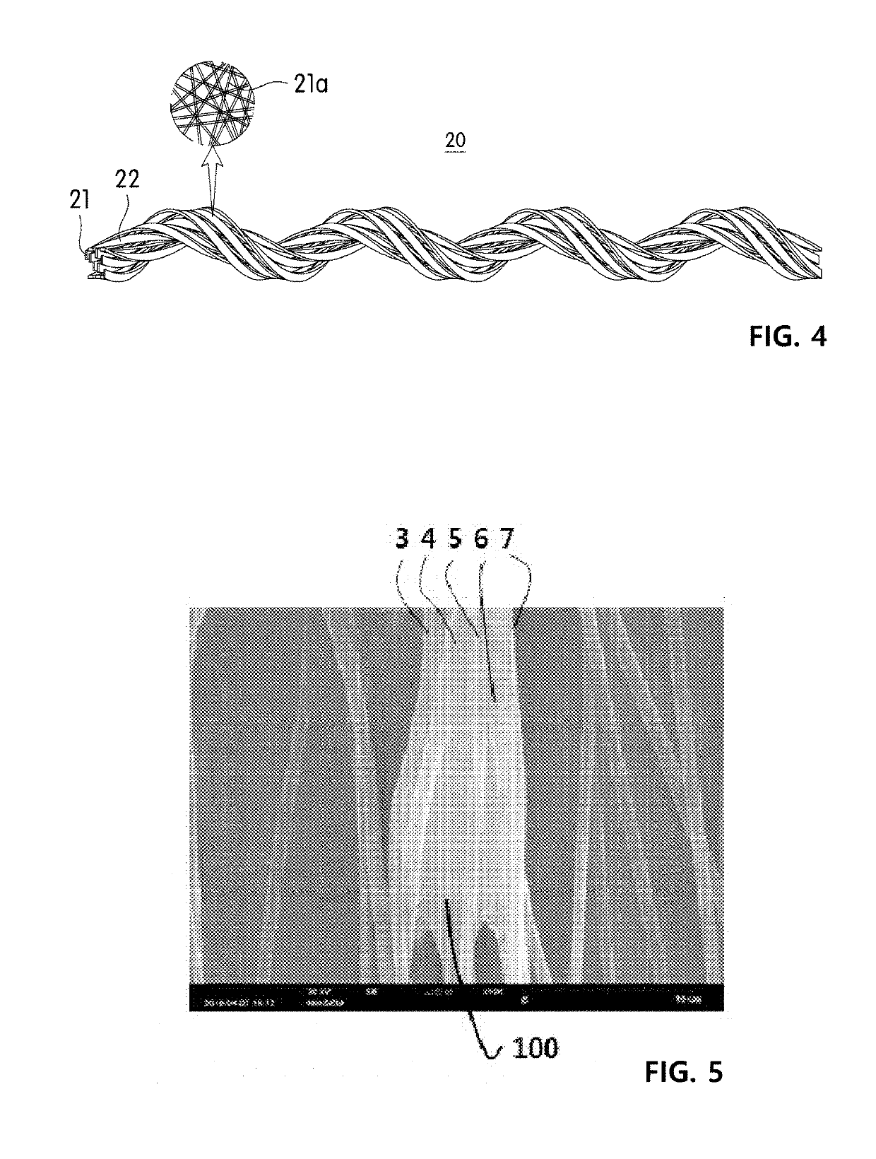
[0083]A plurality of strands of the yarn for a cell culture scaffold produced in Example 1 were arranged parallel and fixed on a well plate for cell culture. Fibroblasts (HS27) were loaded into the well plate including the yarn, and proliferated in a 10% complete medium at 37° C. for 2 days. Here, the 10% complete medium was prepared by mixing Ham's F12 medium with Dulbecco's Modified Eagle Medium (DMEM) at a volume ratio of 1:1.5, and adding 7 vol % of fetal bovine serum, 65 U / mL of penicillin and 65 μg / mL of streptomycin. Afterward, an SEM image of the proliferated fibroblasts was taken and is shown in FIG. 5, and a confocal microscope image of the fibroblasts taken after DAPI staining is shown in FIG. 6.

[0084]Referring to FIGS. 5 and 6, it can be confirmed that the fibroblasts are cultured in contact with an open space between the plurality of twisted fiber strands by partial untwisting, and it can be expected that, when fibroblasts are adhered onto different spaces shown in FIG....


PUM

PropertyMeasurementUnit
Linear densityaaaaaaaaaa
Weightaaaaaaaaaa

Abstract

Provided is yarn for a cell culture scaffold. The yarn includes ply-twisted fiber strands, and to prevent density-dependent inhibition of cultured cells and increase a cell-contacting specific surface area, at least a part of the plurality of twisted fiber strands are untwisted such that an open space is formed between the fibers. A cell proliferation rate and cell viability may be increased by creating microenvironments suitable for migration, proliferation and differentiation of the cultured cells using the yarn. A large quantity of cells may be simultaneously cultured by creating a cell proliferation space as large as possible in a scaffold space having a limited cell proliferation space, and cell proliferation may be steadily maintained by preventing the inhibition of cell proliferation due to intercellular contact. The cells cultured may be cultured to have a shape/structure suitable for application to an in vitro experiment model or implantation into an animal body.

Description

TECHNICAL FIELD[0001]The present invention relates to yarn for a cell culture scaffold, and more particularly, to yarn for a cell culture scaffold which is improved in cell viability by creating microenvironments suitable for adhesion, migration, proliferation and differentiation of cultured cells, allows cells to be three-dimensionally proliferated, prevents density-dependent inhibition by contact between cells proliferated in a limited space according to cell proliferation and increases a specific surface area with which cells can contact, ply yarn including the same, and a fabric including the same.BACKGROUND ART[0002]Recently, according to expansion of the use of cultured cells in disease treatment, interest and research on cell culture are increasing. Cell culture is a technique for collecting cells from a living organism and culturing the cells outside the living organism, and the cultured cells may be used in treatment of various diseases through differentiation into various ...

Claims


Application Information

Patent Timeline
IPC IPC(8): A61L27/38D02G1/02C12N5/0735C12N5/00C12N5/079C12N5/077
CPCA61L27/3834D02G1/02C12N5/0606C12N5/0062C12N5/0618C12N5/0658C12N5/0653C12N5/0654C12N5/0068C12N2513/00C12N2533/30C12N2535/00D02G3/448A61L27/16A61L27/18A61L27/54A61L2300/412A61L27/3804A61L27/3895A61L27/44D02G3/26D10B2509/00
Inventor SEO, IN YONGJANG, SEON HOKOO, SONG HEEKIM, CHANLEE, SEOUNG HOON
Owner AMOLIFESCI CO LTD
Why Eureka
Try Eureka