Nanofiber-hydrogel composites for enhanced soft tissue replacement and regeneration

Pending Publication Date: 2020-03-05
THE JOHN HOPKINS UNIV SCHOOL OF MEDICINE
View PDF5 Cites 4 Cited by
  • Summary
  • Abstract
  • Description
  • Claims
  • Application Information

AI Technical Summary

Benefits of technology

[0012]The compositions and methods in the following disclosure have been designed to addresses this need by using compositions comprising fiber-hydrogel composites

Problems solved by technology

Soft tissue defects resulting from trauma, oncologic resection, or congenital malformation are difficult to treat by conventional means.
Current therapies, including tissue rearrangements or tissue transfer, cause donor site defects.
Other therapies, such as prosthetic implants, lead to fibrosis and encapsulation.
Existing strategies to promote tissue ingrowth are also inadequate for the treatment of soft tissue defects.
Current acellular matrices result in flat, fibrotic sheets of tissue rather than the soft, three-dimensional tissue required for ideal reconstructions.
Finally, while fat grafting can restore soft tissue defects, its wider use is hampered by variable graft survival and limited volumes of restoration.
While several hydrogels have shown some

Method used


Image

  • Nanofiber-hydrogel composites for enhanced soft tissue replacement and regeneration
  • Nanofiber-hydrogel composites for enhanced soft tissue replacement and regeneration
  • Nanofiber-hydrogel composites for enhanced soft tissue replacement and regeneration

Examples


example 1

orming Composite with Reduced Inflammation Profiles

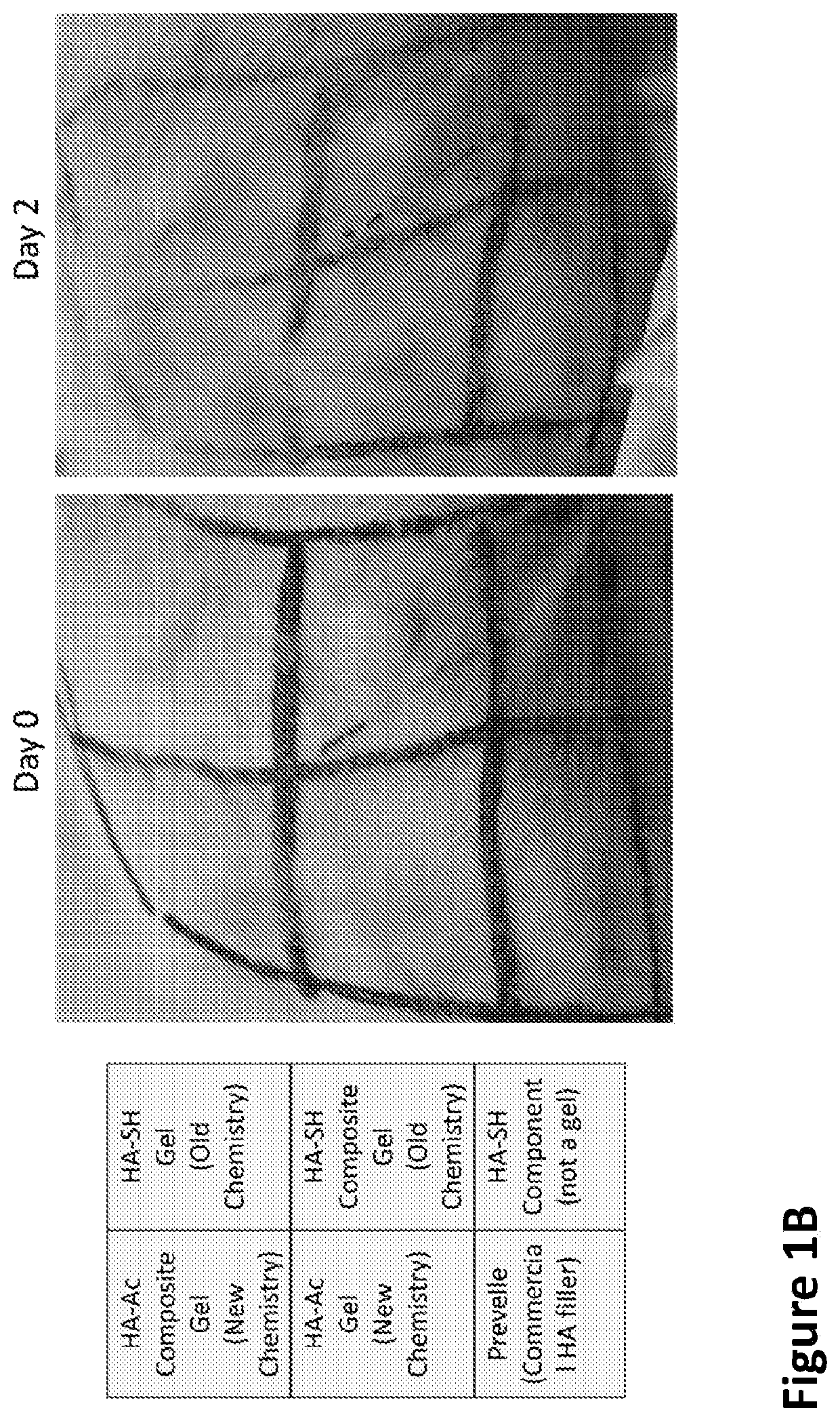
[0240]An in situ-forming composite was developed comprising 5 mg / mL of thiolated HA (HA-SH), 10 mg / mL of polycaprolactone (PCL) fibers and the concentration of PEGDA set to match the thiol concentration 1:1 with the combined acrylate and maleimide concentrations (5 mg / mL). The components were mixed together to react approximately 30 minutes before surgery in order to begin gelation, with the bulk of the gelation being completed in situ.

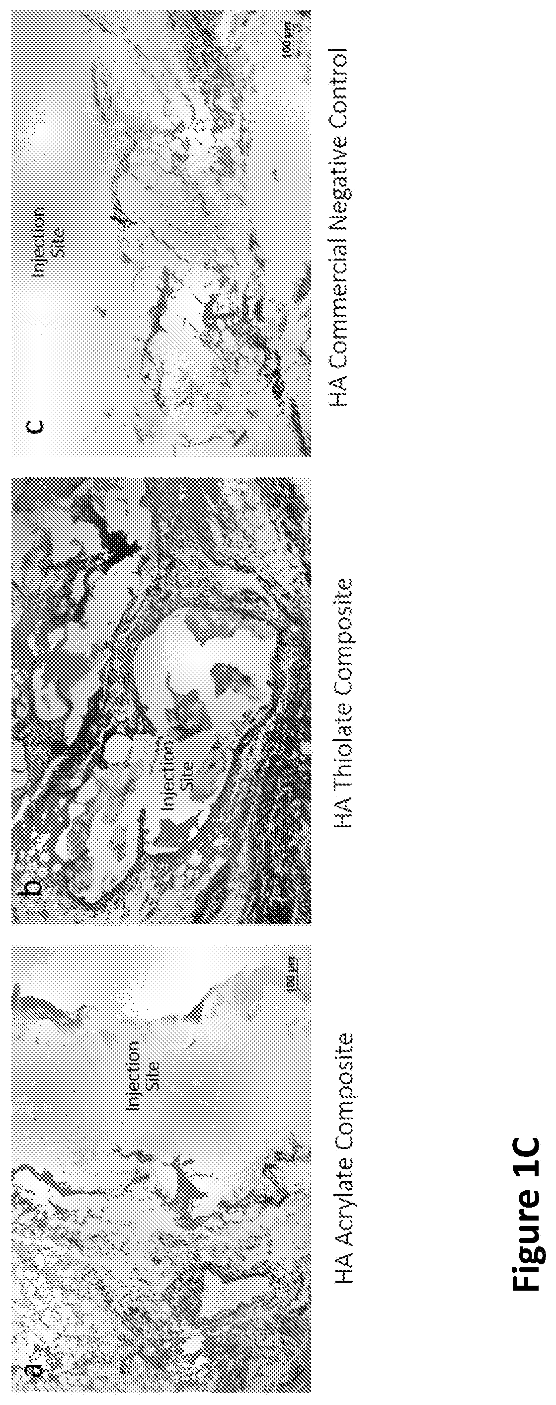
[0241]While gelation success was achieved in vitro and in animals (rodents and rabbits by subcutaneous (s.c.) injection), the chemistry preparation utilizing the thiolated-HA caused short-term moderate inflammation when injected in the subcutaneous rabbit model. In order to produce a composite formulation with reduced inflammation, the reactive groups between the HA and PEG were reversed, keeping an earlier formulation comprising a fiber-maleimide component.

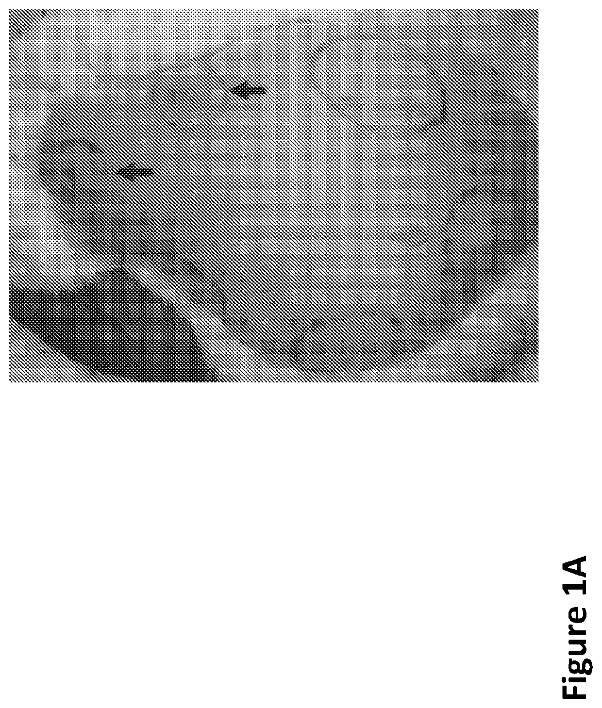
[0242]As shown in FIG. 1A, the upward arrows...

example 3

emical Characterization of Composite Beads

Determination of Size Distribution

[0251]Diameters of composite beads were measured along the longest axis of the particles under a confocal microscope image. The analysis were performed counting 51 particles. Histogram of the particles (FIG. 2G) gives the average bead size as 209.41±62.27 FIG. 2H demonstrates confocal microscope images of beads of size ˜75 μm, ˜150 μm and −200 μm by measuring the longest axis within the particle.

[0252]Further characterized bead size distribution is performed using image analysis program that will use edge detection for more consistent measurements.

[0253]In some embodiments, different mesh size sieves used to process the bulk composite can yield to different histograms for the bead sizes.

[0254]In an alternative embodiment, SEM (Scanning Electron Microscopy) is used to image composite beads. Staining of the hydrogel or fibers may be required during the imaging process.

Assessing Injectability Based on Size of t...

example 8

Assessment with MRI in Rat Model

[0294]The fully-reacted, particulated formulation of the LS series of the composite beads described above shows superior swelling properties when compared to commercial subdermal fillers such as Juvedérm. Specifically, the significant post-procedure swelling observed with Juvedérm injection in the rodent MRI model is not seen with the LS composite, enabling more of a “what you see is what you get” appearance that is desired by clinicians. Continued optimization of HA concentrations, fiber loading, and crosslinking for the current particulated composite form is ongoing and are expected to restore comparable durability to existing commercial standards while retaining enhanced volumization, lessened swelling, and more natural feel. The degree of swelling has been characterized and plotted in FIG. 6A-C.

[0295]Tan δ quantifies the balance between energy loss and storage. A higher Tan δ indicates more liquid-like properties, whereas lower tan δ suggests more...


PUM

PropertyMeasurementUnit
Lengthaaaaaaaaaa
Lengthaaaaaaaaaa
Lengthaaaaaaaaaa

Abstract

A composite material can include a gel and at least one nanostructure disposed within the gel. A method for healing a soft tissue defect can include applying a composite material to a soft tissue defect, wherein the composite material includes a gel and a nanostructure disposed within the gel. A method for manufacturing a composite material for use in healing soft tissue defects can include providing a gel and disposing nanofibers within the gel.

Description

CROSS-REFERENCES TO RELATED APPLICATIONS[0001]This application is a Continuation Application of PCT / US19 / 31638, filed on May 9, 2019 which claims the benefit of U.S. Provisional Application 62 / 669,307 filed May 9, 2018, which are incorporated by reference herein in their entirety.GOVERNMENT SUPPORT[0002]This invention was made with government support under grant no. 1R21NS085714 awarded by the U.S. National Institutes of Health and grant no. DMR1410240 awarded by the National Science Foundation. The government has certain rights in the invention.SEQUENCE LISTING[0003]The instant application contains a Sequence Listing which has been submitted electronically in ASCII format and is hereby incorporated by reference in its entirety. Said ASCII copy, created on Jul. 17, 2019, is named 048317-556001US_SL.txt and is 802 bytes in size.BACKGROUNDField[0004]The present disclosure relates to composite materials and methods that restore lost soft tissue volume while promoting soft tissue regene...

Claims


Application Information

Patent Timeline
IPC IPC(8): A61L27/56A61L27/48A61L27/54
CPCA61L27/54A61L27/48A61L2430/34A61L27/56C08L5/08A61L27/52A61L27/20A61L2400/06A61L2400/12A61L2300/236A61L2300/62A61L27/3633
Inventor MARTIN, RUSSELLMAO, HAI-QUANREDDY, SASHANKCOLBERT, KEVIN
Owner THE JOHN HOPKINS UNIV SCHOOL OF MEDICINE
Why Eureka
Try Eureka