Mitochondrial treatment of organs for transplantation

a technology of mitochondria and organs, applied in the field of mitochondria, can solve the problems of no known and approved treatments or therapies that involve the treatment of cells, tissues, organs with exogenous mitochondria, cell death by apoptosis, etc., and achieve the effects of improving the performance of implanted tissue, minimizing damage to an organ, and improving the function of a lung

Pending Publication Date: 2021-01-14
UNITED THERAPEUTICS CORP
View PDF6 Cites 2 Cited by
  • Summary
  • Abstract
  • Description
  • Claims
  • Application Information

AI Technical Summary

Benefits of technology

[0010]The present disclosure provides methods of organ transplantation comprising delivering isolated mitochondria to an organ intended for transplantation. In another embodiment, the disclosure provides methods of improving the performance of an implanted tissue or transplanted organ in a subject comprising delivering isolated mitochondria to a tissue or organ before, during, or after implantation or transplantation of the tissue or organ, where the tissue or organ is a donor tissue, donor organ, engineered tissue, or engineered organ. In another embodiment, the disclosure provides methods of improving the function of a lung during ex vivo lung perfusion (EVLP) comprising: (i) delivering isolated mitochondria to a lung, and (ii) performing EVLP on the lung in a chamber or vessel by perfusing the lung with a perfusate solution from a reservoir. In another embodiment, the disclosure provides methods for minimizing damage to an organ ex vivo due to cold ischemia during transportation, shipment, or storage comprising: delivering isolated mitochondria to the organ 0-24 hours before cold ischemia, during cold ischemia, or 0-24 hours after cold ischemia, wherein cells of the organ treated with the isolated mitochondria have at least 5% improvement in mitochondrial function in comparison to cells of a corresponding organ not treated with the isolated mitochondria, and wherein the improved mitochondrial function is increased oxygen consumption and / or increased ATP synthesis.
[0011]In another embodiment, the disclosure provides methods for improving the function of an engineered organ or tissue comprising: (i) preparing an organ or tissue scaffold comprising one or more extracellular matrix components, (ii) populating the organ or tissue scaffold in a bioreactor, chamber, or vessel with populating cells to produce an engineered organ or tissue, and (iii) delivering isolated mitochondria to the engineered organ or tissue. In another embodiment, the disclosure provides methods for improving the function of an engineered organ or tissue comprising: (i) preparing an organ or tissue scaffold comprising one or more extracellular matrix components, and (ii) populating the organ or tissue scaffold in a bioreactor, chamber, or vessel with the populating cells treated with isolated mitochondria to produce an engineered organ or tissue. In another embodiment, the disclosure provides methods for improving the function of an engineered organ or tissue comprising: (i) preparing an organ or tissue scaffold comprising one or more extracellular matrix components, (ii) infusing the organ or tissue scaffold with the isolated mitochondria, and (iii) populating the infused organ or tissue scaffold in a bioreactor, chamber, or vessel with populating cells to produce an engineered organ or tissue.
[0012]In another embodiment, the disclosure provides methods for improving the function of an engineered lung comprising: (i) repopulating a decellularized scaffold lung in a bioreactor, chamber, or vessel with repopulating cells to produce an engineered lung, and (ii) delivering isolated mitochondria to the engineered lung. In another embodiment, the disclosure provides methods for improving the function of an engineered lung comprising: (i) delivering isolated mitochondria to repopulating cells, and (ii) repopulating a decellularized scaffold lung in a bioreactor, chamber, or vessel with the repopulating cells treated with the isolated mitochondria to produce an engineered lung.
[0013]In another embodiment, the disclosure provides methods for improving the function of an engineered kidney comprising: (i) repopulating a decellularized scaffold kidney in a bioreactor, chamber, or vessel with repopulating cells to produce an engineered kidney, and (ii) delivering isolated mitochondria to the engineered kidney. In another embodiment, the disclosure provides methods for improving the function of an engineered kidney comprising: (i) delivering isolated mitochondria to repopulating cells, and (ii) repopulating a decellularized scaffold kidney in a bioreactor, chamber, or vessel with the repopulating cells treated with the isolated mitochondria to produce an engineered kidney.
[0020]In another embodiment, the disclosure provides methods for improving the cold transportation, cold shipment, or cold storage of isolated cells comprising delivering isolated mitochondria to the isolated cells before, during, or after cold transportation, cold shipment, or cold storage, wherein the cells treated with the isolated mitochondria have at least 5% improvement in viability in comparison to corresponding cells not treated with the isolated mitochondria. In another embodiment, the disclosure provides methods for cryopreservation of isolated mitochondria comprising freezing isolated mitochondria in a freezing buffer comprising a cryprotectant. In another embodiment, the disclosure provides methods for long-term storage of isolated mitochondria comprising (i) isolating mitochondria from cells or tissue, (ii) suspending the isolated mitochondria in a cold storage buffer, (iii) freezing the isolated mitochondria at a temperature from about −70° C. to about −100° C., and (iv) maintaining the frozen isolated mitochondria at a temperature from about −70° C. to about −100° C. for 24 hours or longer. The storage period can be at least 24 hours, at least one week, at least four weeks, at least three months, at least six months, at least 9 months, or at least 1 year.

Problems solved by technology

Disruption of the outer mitochondrial membrane results in the leaking of mitochondrial proteins into the cytosol, which triggers cell death by apoptosis.
Currently, however, there are no known and approved treatments or therapies that involve the treatment of cells, tissue, or organs with exogenous mitochondria, such as porcine mitochondria or exogenous human mitochondria (i.e., mitochondria isolated from a first human subject used to treat the cells, tissue, or organs of a second human subject).

Method used


Image

  • Mitochondrial treatment of organs for transplantation
  • Mitochondrial treatment of organs for transplantation
  • Mitochondrial treatment of organs for transplantation

Examples


example 1

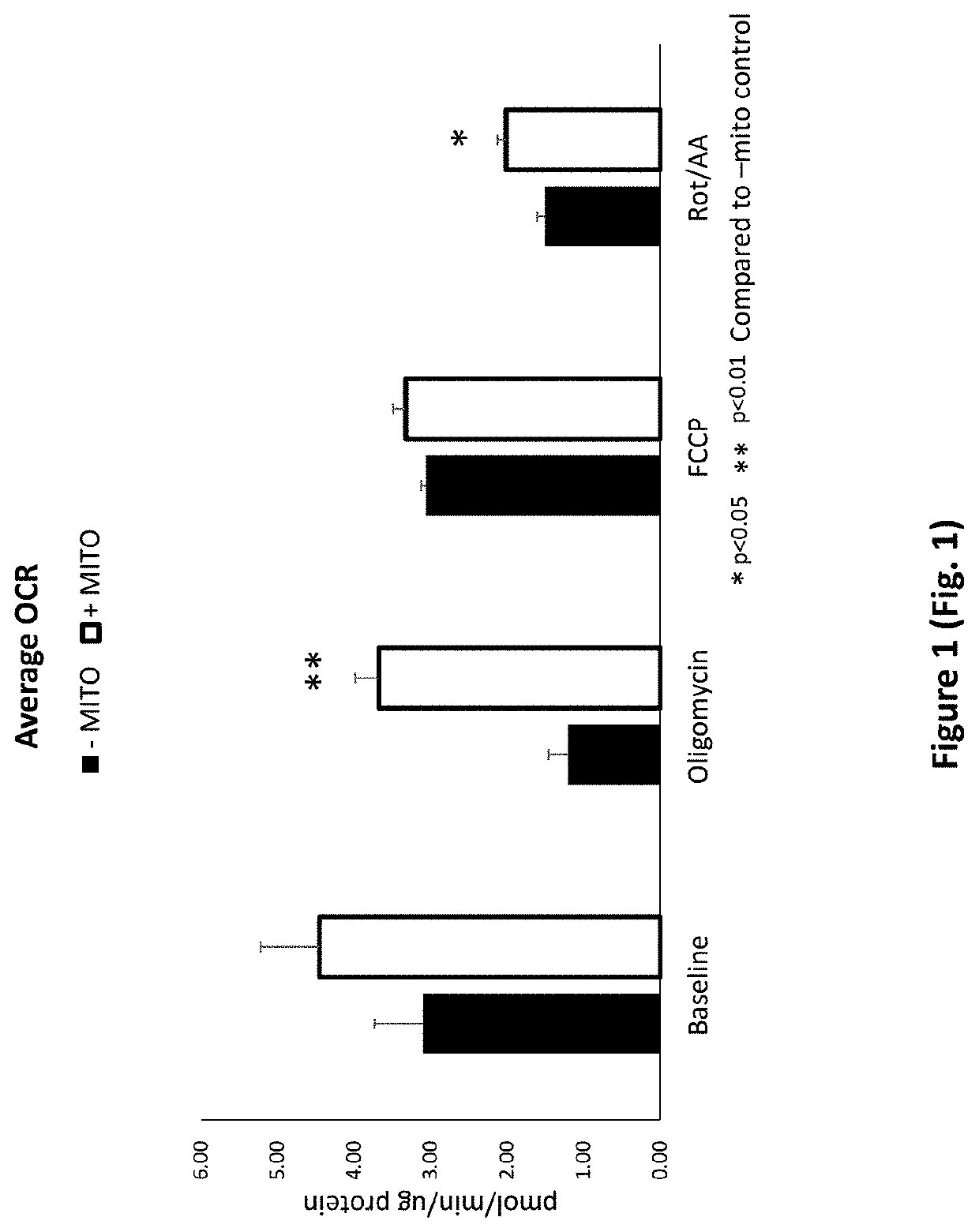
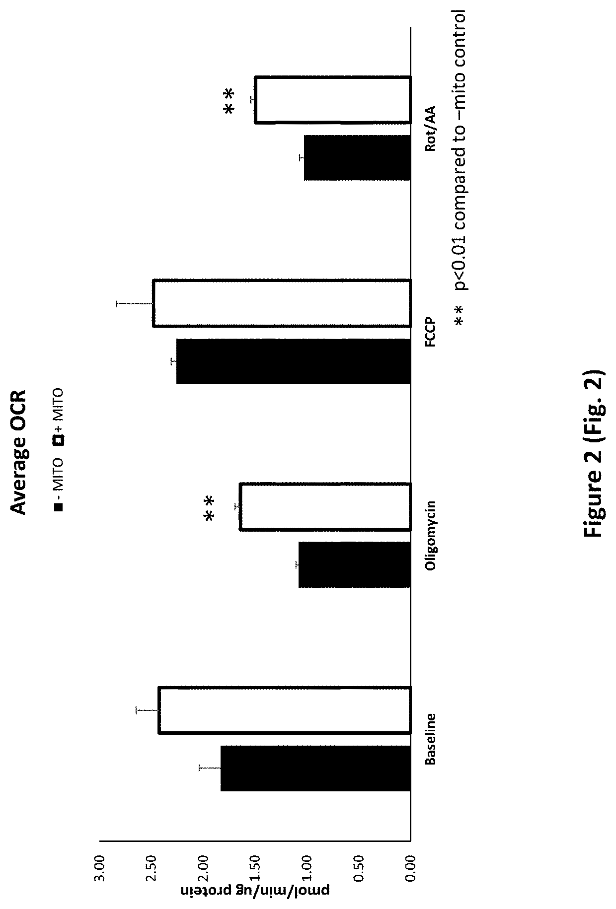
Treatment of Cells with Porcine Mitochondria Improves Oxygen Consumption Rate after Acute and Chronic Cold Exposure

[0296]To isolate porcine mitochondria, the entire left ventricle was removed from a freshly excised pig heart and placed in ice cold washing media (300 mM sucrose, 1 mM EGTA, 10 mM HEPES (pH 7.4)) for transport. A one inch square piece of tissue was cut from the left ventricle and transferred to a pre-chilled 50 ml conical tube containing 20 mL ice cold Trehalose buffer. The tissue sample was minced to obtain sample pieces of approximately 1-2 mm in size. The sample was enzymatically digested in a subtilisin A solution (5 mg / ml subtilisin A in 250 μl of Trehalose buffer) on ice for 10 minutes and homogenized using a Potter-Elvehjem pattern tissue homogenizer (3-7 passes). The sample was then passed through gauze into a 50 ml conical tube. The sample was centrifuged (10 minutes at 4° C. at 500 g) and the supernatant was decanted into a fresh 50 mL conical tube. The sampl...

example 2

Treatment of Cells with Porcine Mitochondria During Cold Recovery and Cold Exposure Alters the Expression of Genes Associated with Inflammation, the Innate Immune Response, and Cell Stress

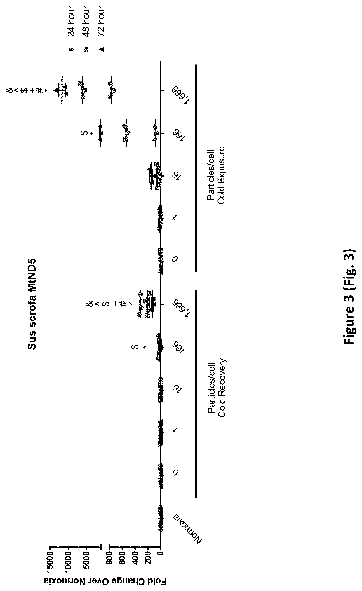
[0303]NF-κB is a transcription factor known to upregulate pro-inflammatory gene expression. The effects of porcine mitochondria treatment on NF-κB gene expression in HPAEC under cold exposure and cold recovery conditions was evaluated by qRT-PCR. As shown in FIG. 5, porcine mitochondria treatment of HPAEC reduces NF-κB expression in cold recovery at 24 hours. HPAEC were treated, cultured under cold recovery or cold exposure conditions, and harvested at 24-hour, 48-hour, or 72-hour time points as described above for FIG. 3. In the cold recovery condition, untreated control HPAEC demonstrated an 83% increase in NF-κB expression at 24 hours compared to normothermia controls. Porcine mitochondria treatment trended to decrease the NF-κB expression compared to untreated cold-recovery control HPAEC, with ...

example 3

Treatment of Cells with Porcine Mitochondria Under Hypoxic Conditions Decreases Secretion of Pro-Inflammatory Gene Products

[0306]To evaluate the effects of porcine mitochondria treatment on human endothelial cells under hypoxic conditions, HPAEC were cultured at normoxia or hypoxia (1% O2) for 24 hours prior to porcine mitochondria treatment. After porcine mitochondria treatment, HPAEC were placed back in their respective conditions, normoxia or hypoxia. 300 μL cell culture media was then collected at 24 hours, 48 hours, or 72 hours and placed in a sterile 1.5 mL Eppendorf tube at the appropriate time point (24 hours, 48 hours, or 72 hours). The tubes were spun down for 10 minutes at 4° C. at 2,000 rpm. The supernatant (270 μL) was collected and placed in a fresh, sterile 1.5 mL Eppendorf tube. These samples were immediately stored at −80° C. until analysis by inflammatory cytokine array. Secreted pro-inflammatory gene products were measured in the cell culture media using an inflam...


PUM

PropertyMeasurementUnit
temperatureaaaaaaaaaa
timeaaaaaaaaaa
timeaaaaaaaaaa

Abstract

Methods and compositions relating to isolated mitochondria are disclosed. For example, cells, tissues, or organs can be treated with isolated mitochondria, such as porcine mitochondria, to improve mitochondrial function in the cell, tissue, or organ. The improvements to mitochondrial function include increased oxygen consumption and increased ATP synthesis. Such methods and compositions are useful for cell therapy; organ and tissue transplantation; organ and tissue engineering; and cold storage or shipment of harvested organs, tissues, and cells.

Description

CROSS-REFERENCE TO RELATED APPLICATIONS[0001]This application claims priority to U.S. Provisional Application No. 62 / 863,034, filed Jun. 18, 2019.FIELD OF THE INVENTION[0002]This disclosure relates to the use of mitochondria, such as isolated porcine mitochondria or isolated human mitochondria, for improving cell, tissue, and organ function and to the therapeutic use of mitochondria.BACKGROUND OF THE INVENTION[0003]The mitochondrion is a double-membrane-bound organelle in eukaryotic cells that plays a key role in the maintenance and preservation of cellular homeostasis and function. For example, mitochondria supply cellular energy and play a key role in cell signaling, cellular differentiation, cellular apoptosis, cell cycle regulation, and cell growth. Typically, mitochondria supply more than 90% of a cell's ATP requirement.[0004]The mitochondrion is composed of an outer mitochondrial membrane, an inner mitochondrial membrane, an intermembrane space between the outer and inner memb...

Claims


Application Information

Patent Timeline
Patent Type & Authority Applications(United States)
IPC IPC(8): A61K35/12C12N5/071A61P11/00
CPCA61K35/12A61P11/00C12N5/0602A61K35/34A61P13/12A61P9/10A01N1/0226A61L27/3683A61L27/3687A61L27/3604A61L2430/40A61L2430/26A61L27/3633
Inventor PETERSEN, THOMASHOGAN, SARAHILAGAN, ROGERCLOER, CARYN
Owner UNITED THERAPEUTICS CORP
Why Eureka
Try Eureka