Activin receptor type iia variants and methods of use thereof

Active Publication Date: 2021-02-04
KEROS THERAPEUTICS INC
View PDF0 Cites 1 Cited by
  • Summary
  • Abstract
  • Description
  • Claims
  • Application Information

AI Technical Summary

Benefits of technology

[0109]FIG. 3 is graph showing the effect of treatment with ActRIIA/B-Fc on trabecular bone volume fraction in a mouse model of osteoporos

Problems solved by technology

Pathology occurs when these systems fall out of balance, either through downregulation of the anabolic program, upregulation of the catabolic system, or a combination of both, resulting in a net bone loss.
Bone damage can result from a range of root causes, including age- or cancer-related bone loss, genetic con

Method used


Image

  • Activin receptor type iia variants and methods of use thereof
  • Activin receptor type iia variants and methods of use thereof
  • Activin receptor type iia variants and methods of use thereof

Examples


example 1

Evaluation of ActRIIa Variants Binding Affinity by Surface Plasmon Resonance (SPR)

[0175]The Biacore 3000 was used to measure the kinetics of the interactions between the ActRIIa variants and the ligands Activin A, Activin B, growth differentiation factor 11 (GDF11), and BMP-9. ActRIIa variants were transiently expressed in HEK293 cells and purified from the conditioned media using Protein-A Sepharose chromatography. The ActRIIa variants were immobilized on the chip (CM4 or CM5) with capture antibodies (anti-mouse from GEGE) in flow cells 2-4 to ensure proper orientation. Flow cell 1 was used as a reference cell to subtract any nonspecific binding and bulk effects. HBS-EP+ buffer from GE Healthcare™ was used as a running buffer. Each ligand was run in a concentration series at 40 μl / min to avoid mass transport effects. The data was analyzed using Scrubber2 by BioLogic™ Software to calculate the KD of each interaction (Table 5).

TABLE 5Comparison of Act RIIa variant binding affinity (K...

example 2

Effect of Extracellular ActRIIa Variants on Bone Mineral Density

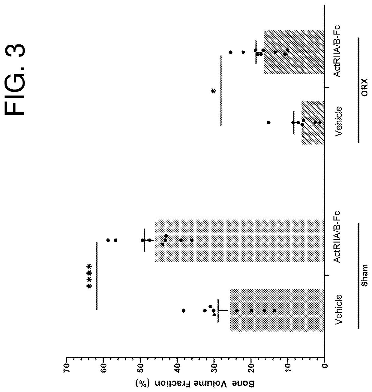
[0176]Adult male C57 / BL6 mice receive either a sham- (SHAM) or castration-surgery (ORX). Both surgery groups are allowed to recover for 14 days post-surgery. All animals are housed in conventional cages with free access to food (regular chow) and water. SHAM and ORX animals are then assigned to either a vehicle-treated group (VEH) or ActRII variant-treated group and receive bi-weekly systemic intraperitoneal administration of vehicle or ActRII variant (10 mg / kg) for 71 d. Body weights are measured twice per week at the time of treatment. Body composition is analyzed at study day 0 then at days 14, 28, 47, and 71 after treatment initiation using the MiniSpec LF50 NMR Analyzer. At study termination date, tissues of interest (muscles, fat depots, and tibias) are surgically removed, weighed, and properly stored for further analysis. At this time, the ORX animals are also examined to confirm complete removal of testes. Corti...

example 3

Effect of Extracellular ActRIIa Variants on Trabecular Bone

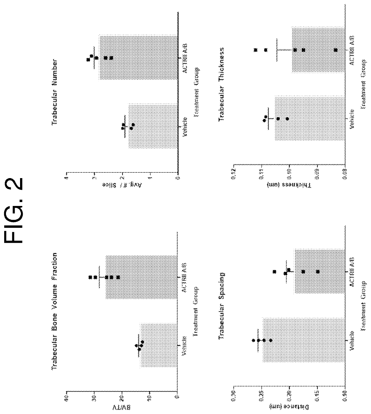
[0177]Eight-week old male C57Bl / 6 mice were dosed intraperitoneally with either vehicle or ActRII A / B (SEQ ID NO: 69) at 20 mg / kg biweekly for four weeks. Upon completion of dosing, mice were micro-CT imaged at high resolution with the PerkinElmer Quantum FX system (10 mm FOV, 3 min scan, 20 μm voxel-size). Tibia ASBMR bone morphometry parameters were measured in AnalyzePro software from a 50-slice region of scan volume selected immediately distal to the proximal tibial growth plate. From this sub-region trabecular bone fraction, trabecular number, trabecular thickness, and trabecular spacing data were calculated (FIG. 2). Treatment with ActRII A / B (SEQ ID NO: 69) resulted in increased trabecular bone volume fraction, trabecular number, and decreased trabecular spacing. These changes to trabecular bone are associated with increased bone strength and reduced fracture risk.


PUM

PropertyMeasurementUnit
Densityaaaaaaaaaa
Gravityaaaaaaaaaa
Strengthaaaaaaaaaa

Abstract

The invention features polypeptides that include an extracellular ActRIIa variant. In some embodiments, a polypeptide of the invention includes an extracellular ActRIIa variant fused to an Fc domain monomer or moiety. The invention also features pharmaceutical compositions and methods of using the polypeptides to treat diseases and conditions involving bone damage, e.g., primary osteoporosis, secondary osteoporosis, osteopenia, osteopetrosis, fracture, bone cancer or cancer metastasis-related bone loss, Paget's disease, renal osteodystrophy, treatment-related bone loss, diet-related bone loss, bone loss associated with the treatment of obesity, low gravity-related bone loss, or immobility-related bone loss.

Description

BACKGROUND OF THE INVENTION[0001]Healthy bone undergoes a constant remodeling that involves both bone breakdown and bone growth. Bone growth is mediated by the osteoblast cell type, whereas the osteoclasts resorb the bone. Pathology occurs when these systems fall out of balance, either through downregulation of the anabolic program, upregulation of the catabolic system, or a combination of both, resulting in a net bone loss. Therefore, controlling the balance in bone remodeling can be useful for promoting the healing of fractures and other damage to bone as well as for treating disorders, such as osteoporosis, that are associated with loss of bone mass and bone mineralization.[0002]Bone damage can result from a range of root causes, including age- or cancer-related bone loss, genetic conditions, adverse side effects of drug treatment, and fracture. The World Health Organization estimates that osteoporosis alone affects 75 million people in the U.S., Europe, and Japan, and is a signi...

Claims


Application Information

Patent Timeline
IPC IPC(8): A61K38/17A61P19/00A61P19/10
CPCA61K38/179A61P19/10A61P19/00C07K14/71A61P19/08A61K38/1796A61K38/1825A61K38/26C07K2319/30A61K2300/00A61K38/18
Inventor LACHEY, JENNIFERSEEHRA, JASBIR S.
Owner KEROS THERAPEUTICS INC
Why Eureka
Try Eureka