Suppression of neointimal formation following vascular surgeru using cdk8 inhibitors
a vascular surgery and inhibitor technology, applied in the field of vascular surgery intervention, can solve the problems of limited long-term efficacy of cabg surgery, failure of cabg treatment, so as to reduce postoperative cardiovascular events, reduce neointimal hyperplasia, and limit the progression of atherosclerosis
- Summary
- Abstract
- Description
- Claims
- Application Information
AI Technical Summary
Benefits of technology
Problems solved by technology
Method used
Image
Examples
example 1
[0056]Male C57BL / 6J mice were purchased from Vital River Laboratory Animal Technology Co. Ltd, Beijing, China, and housed under a 12:12 h light-dark cycle and given free access to food and water. All animal experiments were performed according to National Institutes of Health Guidelines for the Care and Use of Laboratory Animals and approved by the Institutional Animal Care and Usage Committees at Shandong University and the University of South Carolina, USA. The jugular vein transplantation was performed using different male C57BL / 6J mice as donors and recipients. Briefly, 12-wk-old male C57BL / 6J mice were anesthetized with pentobarbital sodium (50 mg / kg body weight, i.p.). The operation was performed under a dissecting microscope (SZ2-ILST, Olympus Corporation, Tokyo, Japan). The right common carotid artery of a male C57BL / 6J mouse was mobilized and divided. A 1-mm cuff with a 1-mm handle (0.65 mm in diameter outside and 0.5 mm inside, F 800 / 200 / 100 / 200...
example 2
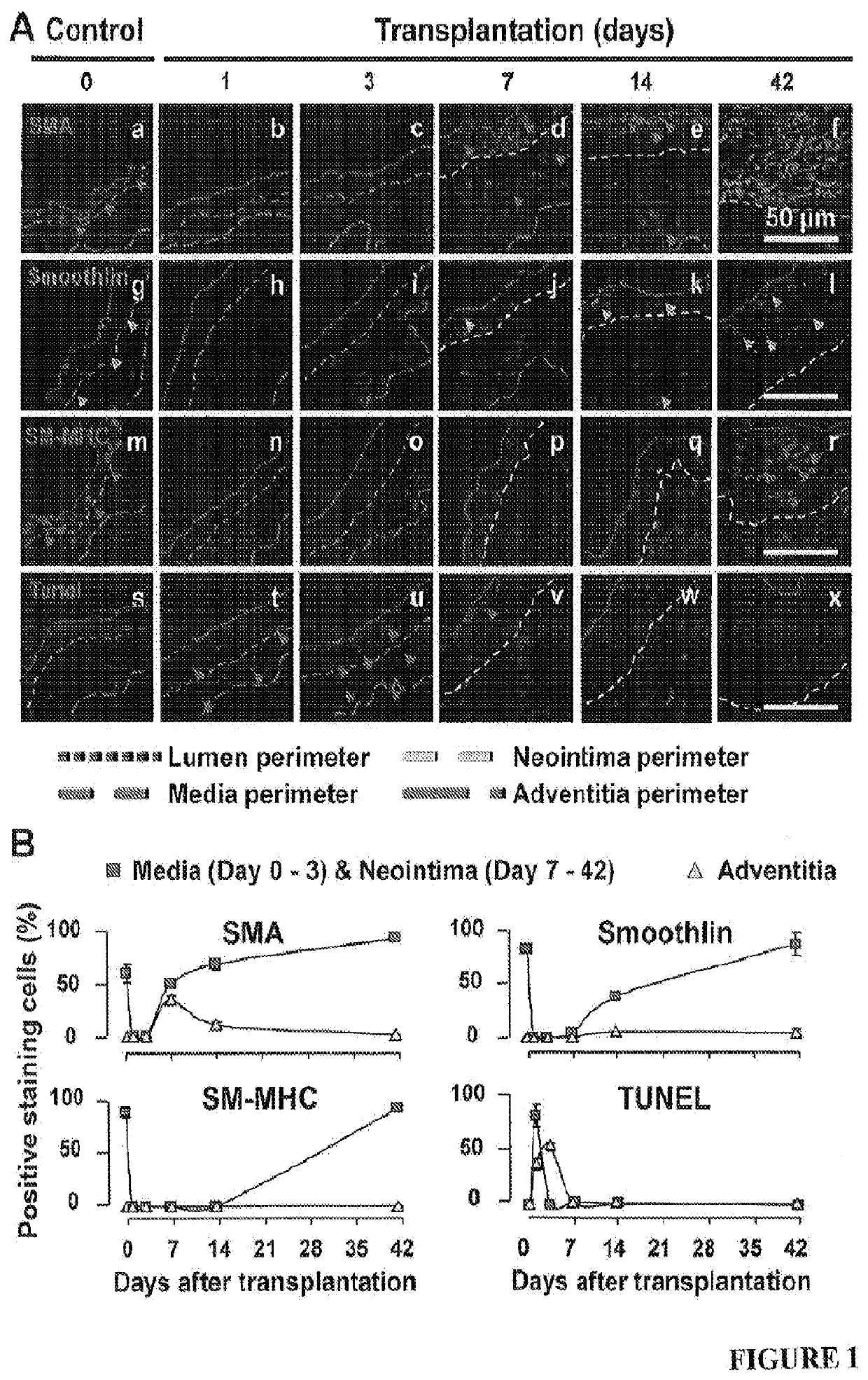
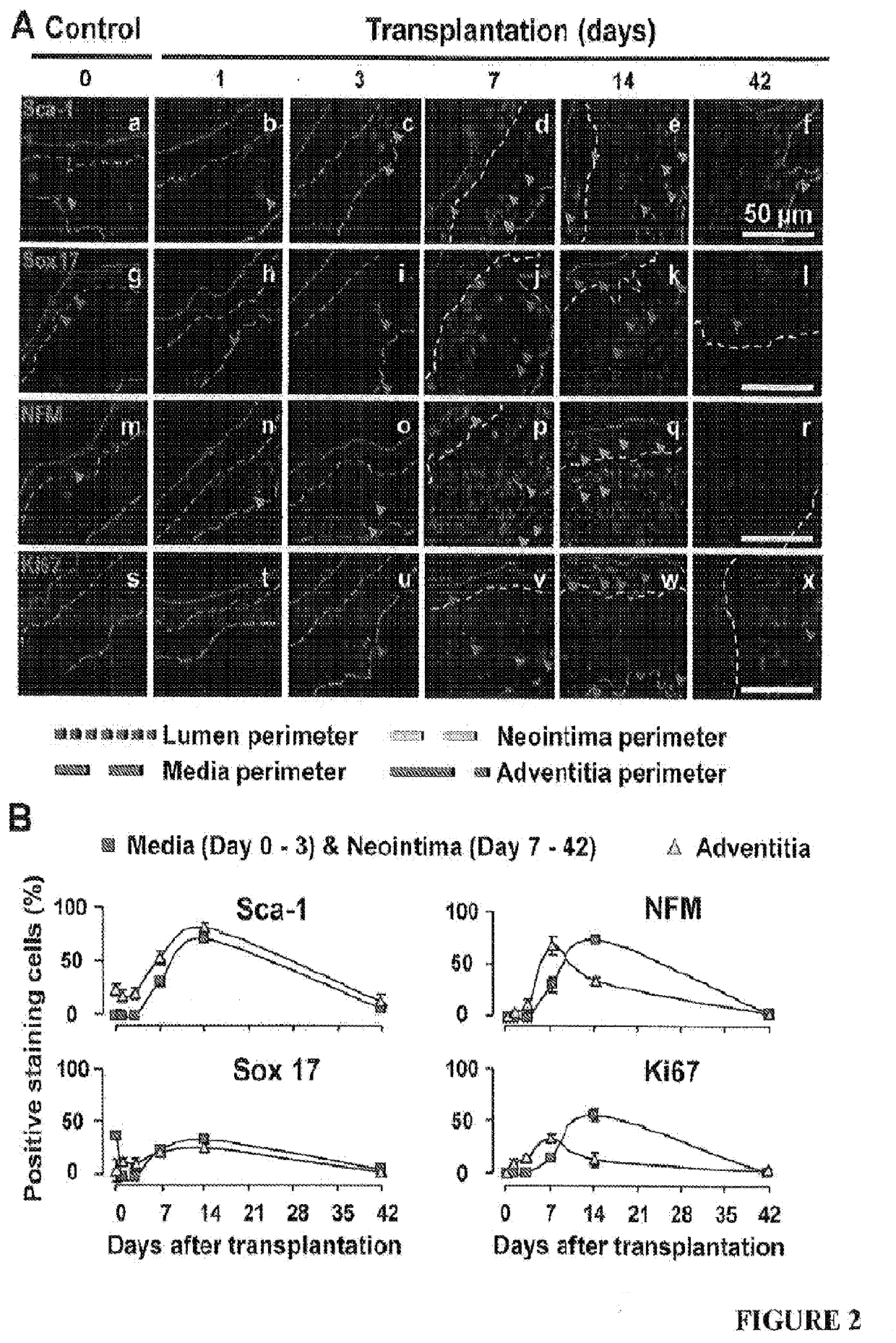
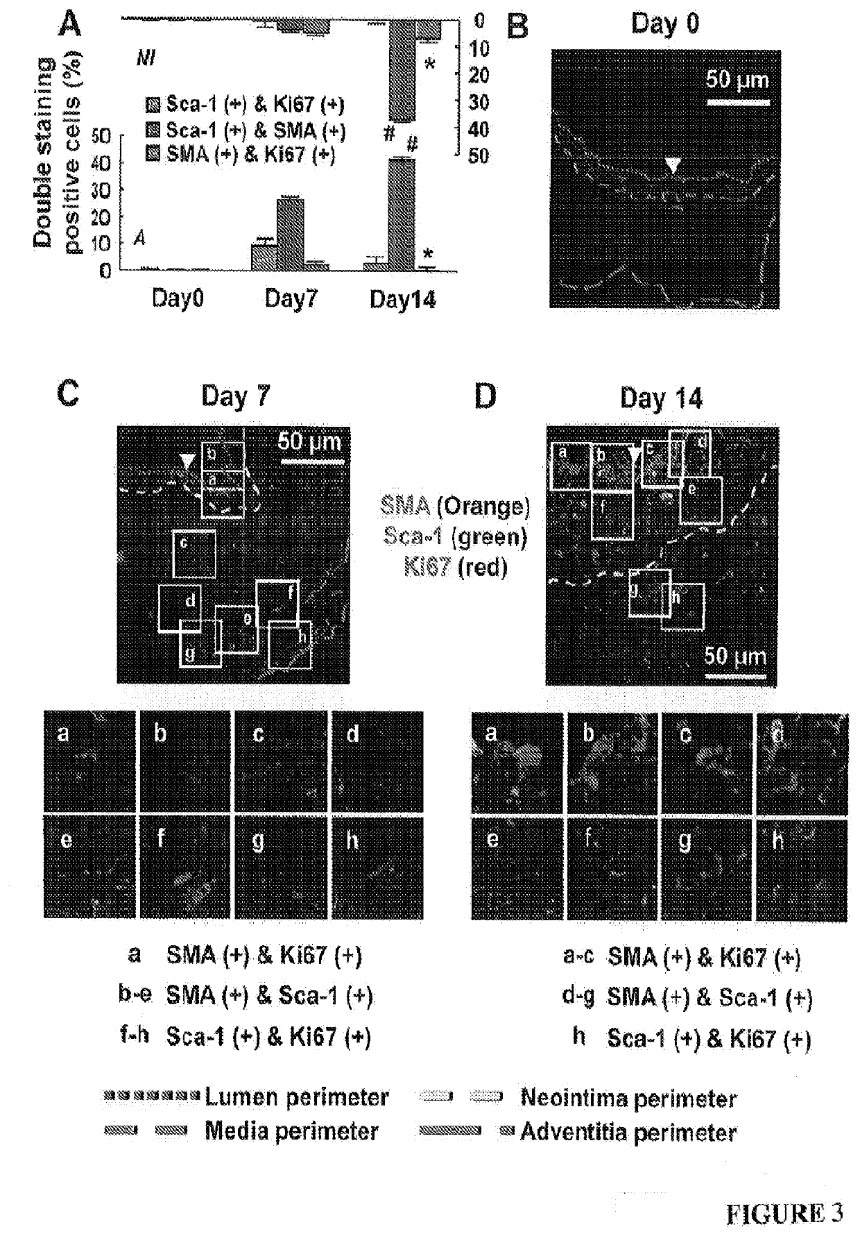
[0060]Mice at 0, 1, 3, 7, 14, 21 and 42 days after transplantation (four randomly chosen mice at each time point) were anesthetized with pentobarbital sodium (50 mg / kg body weight, i.p.) and euthanized by exsanguination. Blood vessels were fixed with 10% formalin under 100 mmHg (13.3 kPa) pressure for 10 min and then washed with by 0.9% NaCl for 5 min. The vein grafts were harvested by cutting the transplanted segments from the native carotid arteries at the two cuff ends. The proximal ends of vein grafts were marked with 8-0 silk ligature. These grafts were further fixed with 4% phosphate-buffered formaldehyde at 4° C. for 24 h, and then embedded in paraffin or OCT for sectioning. All the vein grafts were sectioned from the proximal end.
[0061]From the proximal end, the last section with the cuff tube was counted as #0. Serial cross-sections (5-μm thick per section) were then cut in 5-mm length without exposing the distal end with attached cuff tube. One section from every ...
example 3
[0063]Lumen areas, neointima areas, and neointima thickness were measured as previously described1. In addition, considering the irregularly configured or even closed lumens of vein grafts during the tissue processing, lumen areas were also predicted by a formula: lumen area=πr2 (Supplementary FIG. 1D). By tracking the perimeter length of a lumen which is minimally affected by the tissue processing, r, the radius of a lumen was calculated by a formula: r=perimeter of lumen length / 2n. Similarly, the neointima thickness (ΔNT) was also calculated by a formula: ΔNT=R1−r (Supplementary FIG. 1D). Moreover, we enabled the SMA positive area (SMA area) to be calculated automatically by setting a fluorescent threshold for the immunofluorescent staining images of SMA using Volocity software (PerkinElmer, Waltham, Mass., USA). Accordingly, SMA area thickness, which reflects SMC layer thickness, was calculated by a formula: ΔSMA=R2−r (Supplementary FIG. S1D).
[0064]We reprod...
PUM
 Abstract
Description
Claims
Application Information
 - R&D
- Intellectual Property
- Life Sciences
- Materials
- Tech Scout
- Unparalleled Data Quality
- Higher Quality Content
- 60% Fewer Hallucinations
Browse by: Latest US Patents, China's latest patents, Technical Efficacy Thesaurus, Application Domain, Technology Topic, Popular Technical Reports.
© 2025 PatSnap. All rights reserved.Legal|Privacy policy|Modern Slavery Act Transparency Statement|Sitemap|About US| Contact US: help@patsnap.com


