
Microfluidic chip for biomolecule crystallization

Inactive Publication Date: 2005-02-01
CYTONOMEST
View PDF22 Cites 14 Cited by
  • Summary
  • Abstract
  • Description
  • Claims
  • Application Information

AI Technical Summary

Benefits of technology

The present invention provides for methods and hardware for growing crystals of biological macromolecules using real-time, or dynamic, control of parameters that promote crystal growth from protein solution. Prior crystallization devices available for high throughput screening of crystallization conditions are incapable of addressing at least some of the features listed below. The present invention provides a microfluidic chip for crystallizing proteins or other biomolecules. The system can grow 5-50 micron size, x-ray quality protein crystals from nanogram quantities of protein. The crystallization chip of the invention allows protein crystallization condition studies to be performed when only a few micrograms of a protein are available.
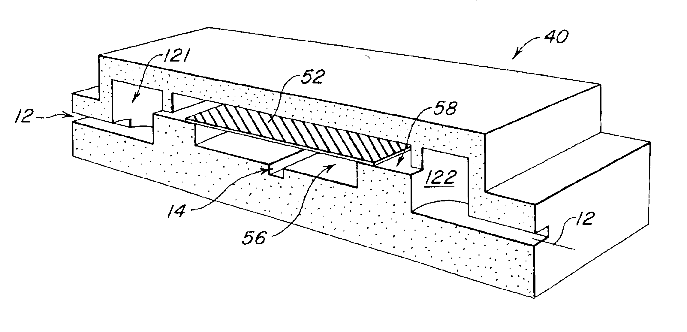
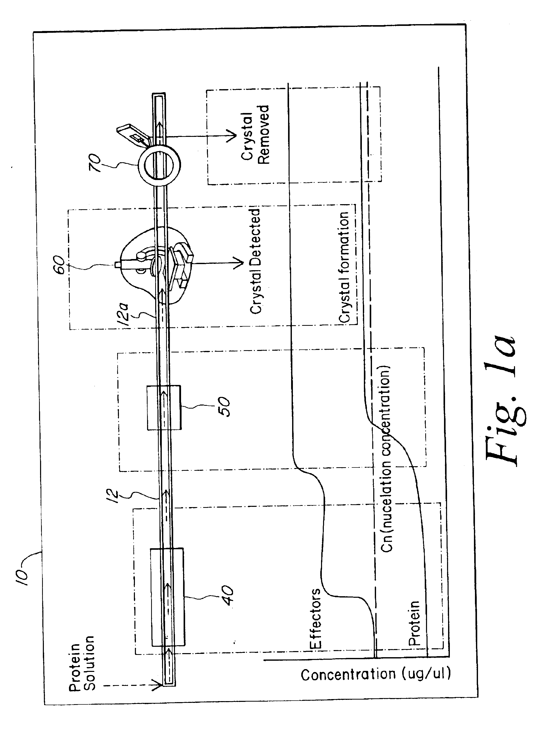
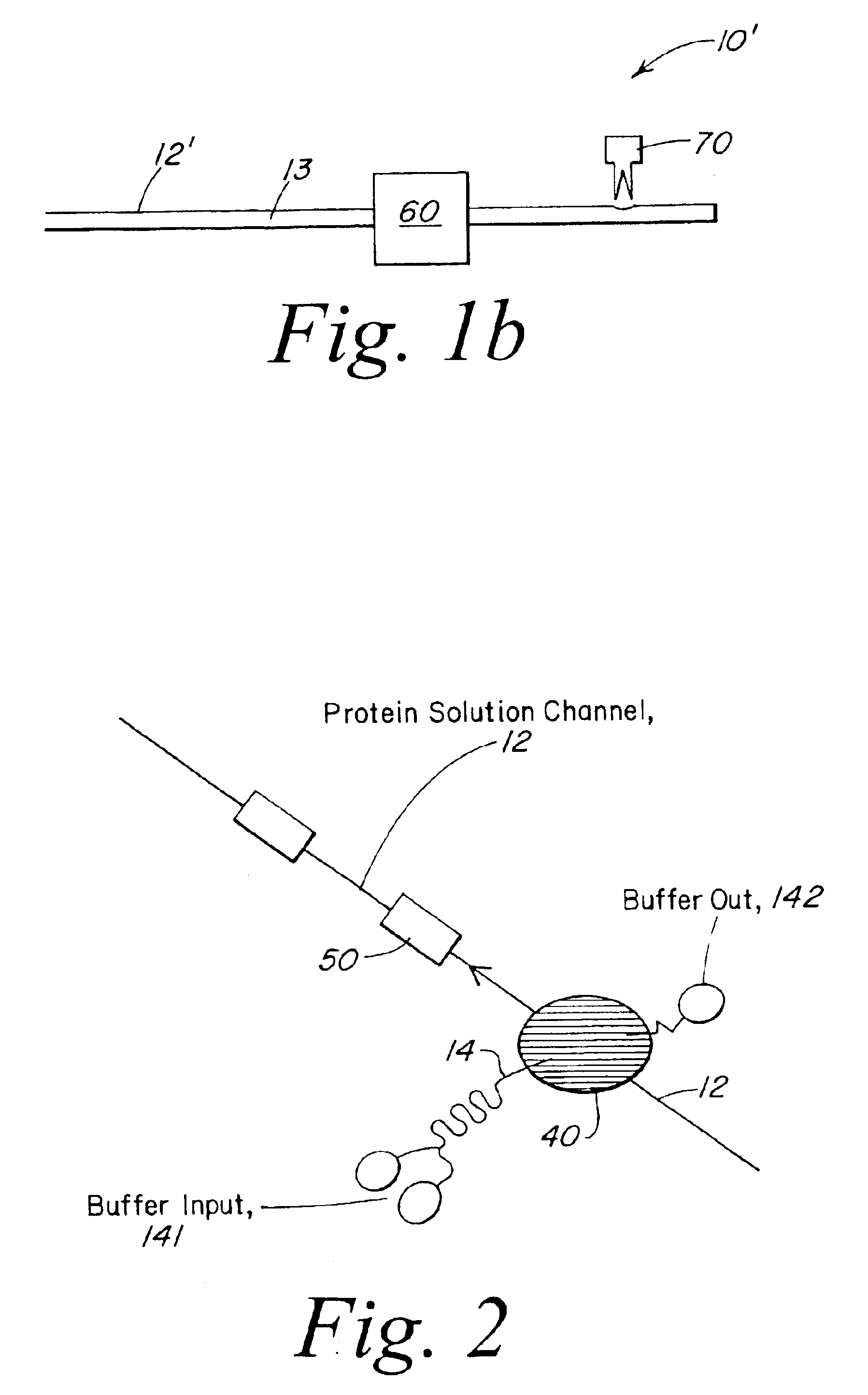
The protein crystallization system of the invention is formed by locating several stations in series along a single flow channel. The system comprises a protein channel for conveying a protein solution containing a protein to be crystallized and an effector station located along the protein channel for selective exchange of solutes between the protein channel and a buffer channel or reservoir by dialysis. A concentration station may also be located along the protein channel for concentrating the protein solution after dialysis. The concentration station may remove water or solvent from the protein solution through a protein impermeable membrane or by evaporation through an aperture in the side wall of the protein channel. The concentration station is followed by a crystallizing region of the protein channel in which crystallization can take place. The protein crystallization system may further include an observation station or region formed along the protein channel. The observation station provides means to monitor each crystallization process, in real time, by polarized light optics to detect presence of crystals or light scattering optics to detect protein aggregation. This allows fine adaptive control of the automation system. A harvesting station may be located downstream from the observation station to provide a means to transfer the crystals from the formation location to the x-ray diffraction apparatus in which crystals are observed to form by optical means. The harvesting station may harvest the crystal by removing an aliquot of liquid containing a crystal from the channel for freezing and x-ray structure analysis.

Problems solved by technology

Prior crystallization devices available for high throughput screening of crystallization conditions are incapable of addressing at least some of the features listed below.

Method used


Image

  • Microfluidic chip for biomolecule crystallization
  • Microfluidic chip for biomolecule crystallization
  • Microfluidic chip for biomolecule crystallization

Examples


Embodiment Construction

The present invention provides a microstructured crystallization system that can be formed on a microfluidic chip. The crystallization system may be used to make single crystals from nanogram quantities of protein or other biomolecule, or to test 600-1000 crystallization conditions from microgram quantities of protein or other biomolecule. The present invention will be described below relative to an illustrative embodiment for crystallizing proteins. Those skilled in the art will appreciate that the present invention may be used to crystallize any desired biomolecule or create co-crystals of any combination of biomolecules and may be implemented in a number of different applications and embodiments. The present invention is not specifically limited in its application to the particular embodiments depicted herein.

The biomolecule crystallization system of the present invention integrates conventional membrane filter technology into a microfluidic chip to perform microdialysis or micro...


PUM


Abstract

A system for crystallizing a protein and other biomolecules comprises a flow channel for conveying a solution containing the biomolecule to be crystallized and one or more substations related to the crystallization process formed along the flow channel. The system operates by flowing a biomolecule solution through a the flow channel, then performing dialysis on a micro-scale to set crystallization conditions. After dialysis, the concentration of the biomolecule solution is changed by removing water or solvent from the biomolecule solution to promote formation of crystals in a segment of the flow channel. Formed crystals may be observed and harvested from the flow channel.

Description

FIELD OF THE INVENTIONThe present invention relates to a microfluidic system and method for crystallizing biomolecules, such as proteins.BACKGROUND OF THE INVENTIONThe understanding of specific properties, functions and three-dimensional structure of proteins is an invaluable asset for understanding protein-ligand interaction and rational drug design. From the basic biochemical standpoint, for example, information on the three-dimensional structure of protein or the like is the basis to understand the functional role of molecules in a biochemical system.In analyzing the structure of proteins, it is generally desirable to grow X-ray diffraction quality crystals from small quantities of a biomolecule. However, structural analysis of protein is currently a labor-intensive, lengthy process that requires significant consumption of expensive macromolecules and reagents. With the advent of robust expression and protein purification systems, high intensity, synchrotron x-ray sources and com...

Claims


Application Information

Patent Timeline
IPC IPC(8): B01L3/00C30B7/00B01L3/06
CPCB01L3/502753C30B7/00B01L3/06C30B29/58B01L3/0248Y10T436/255B01L3/502715B01L3/502723B01L2200/0678B01L2300/0681B01L2300/087B01L2300/1827B01L2400/0487Y10T436/2525Y10T436/25
Inventor GILBERT, JOHNTRIKHA, JAISHREE
Owner CYTONOMEST
Learn More