Universal donor cells

a technology of endothelial cells and donor cells, applied in the field of universal donor cells, can solve the problems of organ transplant rejection and vasculitis, loss of these normal physiological processes, pathological conditions, etc., and achieve the effects of reducing cell surface mhc class ii expression, preventing both complement-mediated lysis and activation of porcine cells, and high degree of endothelial cell expression

Inactive Publication Date: 2005-07-12
OKLAHOMA MEDICAL RES FOUND +1
View PDF7 Cites 35 Cited by
  • Summary
  • Abstract
  • Description
  • Claims
  • Application Information

AI Technical Summary

Benefits of technology

[0042]Examples demonstrate expression of human CD59 in porcine endothelial cells protects the cells from hyperacute rejection by human complement, preventing both complement-mediated lysis and activation of the porcine endothelial cells; stable expression of hCD59 in mouse cells which protects the cells from complement mediated lysis, generation of transgenic mice with a high degree of endothelial cell expression of hCD59 in both systemic and pulmonary circulations, and in capillary, arteriolar, and arterial vasculatures; generation of recombinant mice bearing a deletion of the invariant chain (li) having reduced cell surface MHC class II expression and diminished ability to present exogenous protein antigen; rat capillary endothelial cells engineered to stably express human Apolipoprotein E in three dimensional cultures; and implantation into a rat of the engineered rat capillary endothelial cell network with vascular astomosis to the recipient.

Problems solved by technology

Therefore, loss of endothelial cells results in the loss of these normal physiological processes and ultimately leads to pathological conditions such as coronary artery disease, organ transplant rejection and vasculitis.
In the past, proposals and / or efforts to use endothelial cells for either administration of therapeutics or cell replacement have generally been limited to autologous cells, i.e., cells derived from the organism undergoing treatment.
Alternatively, the patient must be immunosuppressed, which is costly and leaves the patient vulnerable to infection.
This approach has suffered from a number of problems.
For example, it is difficult to harvest healthy endothelial cells from the individual to be treated in significant quantities.
Unfortunately, the cell doubling time for endothelial cells is on the order of at least 24 to 48 hours, leading to time periods on the order of a week or more before sufficient quantities of endothelial cells are available for genetic engineering or cell replacement.
In addition, under normal physiological conditions, the cell doubling time for natural endothelial cells in vivo is also prolonged, making naturally occurring cell replacement in vivo following endothelial cell loss or damage highly inefficient.
This diffusional barrier to the systemic circulation is a considerable impediment to achieving adequate plasma levels of the desired therapeutic protein.
Further, fibroblasts via their excessive production of fibrotic scar tissue, can prove highly detrimental to the host.
Ultimately, endothelial cells were destroyed, resulting in tissue ischemia and necrosis.
Although the use of CD59 does successfully address the problem of hyperacute rejection as a result of complement attack, it does not protect the cell against the overall immune attack of the host organism against foreign endothelial cells.
Historically, attempts to interrupt the T-cell immune response have generally met with limited success.
These strategies have generally resulted in immune responses to the reagents, rather than the desired interruption of T-cell binding.

Method used


Image

  • Universal donor cells
  • Universal donor cells
  • Universal donor cells

Examples


example 1

Expression of Human CD59 in Porcine Endothelial Cells Protects them from Hyperacuto Rejection by Human Complement.

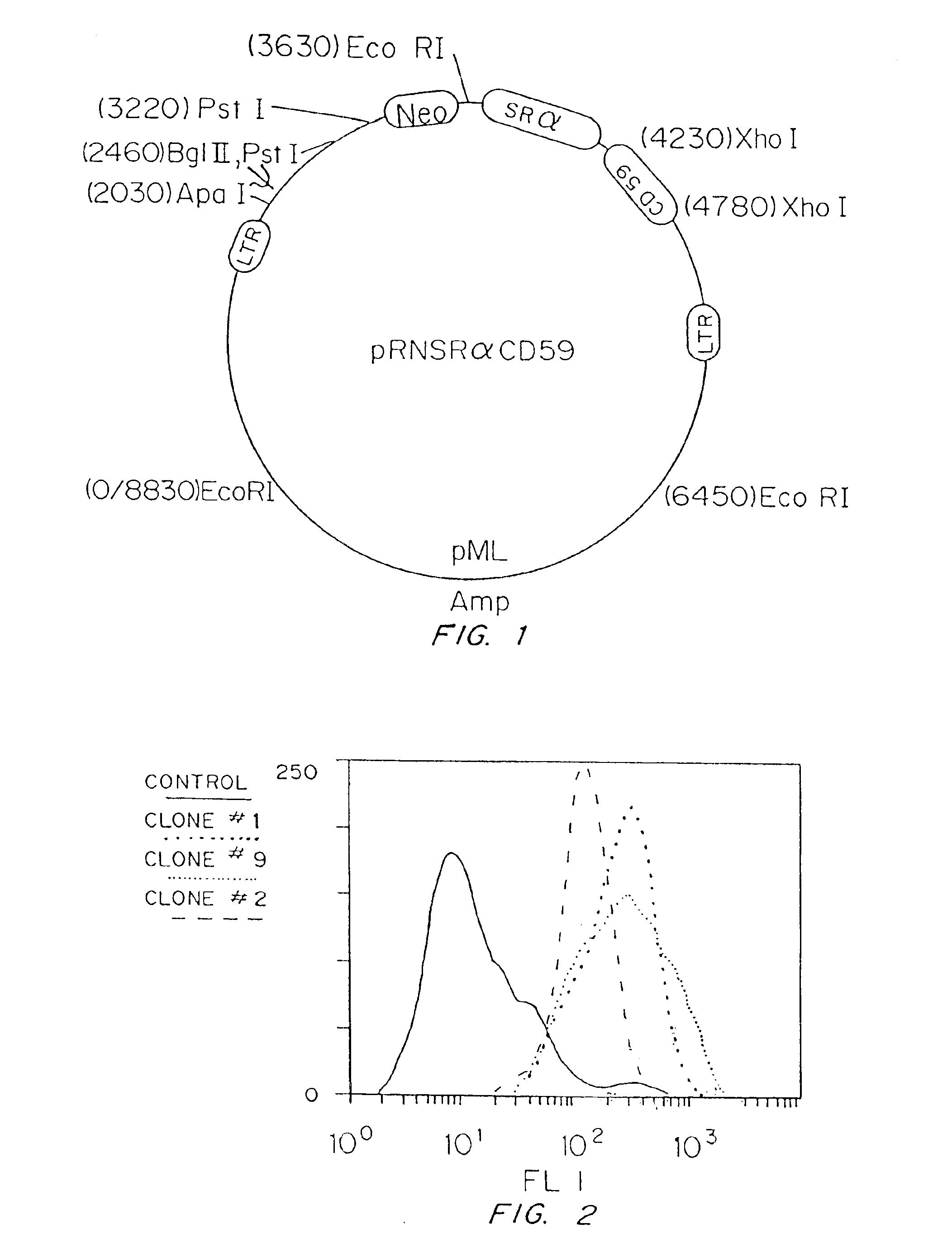
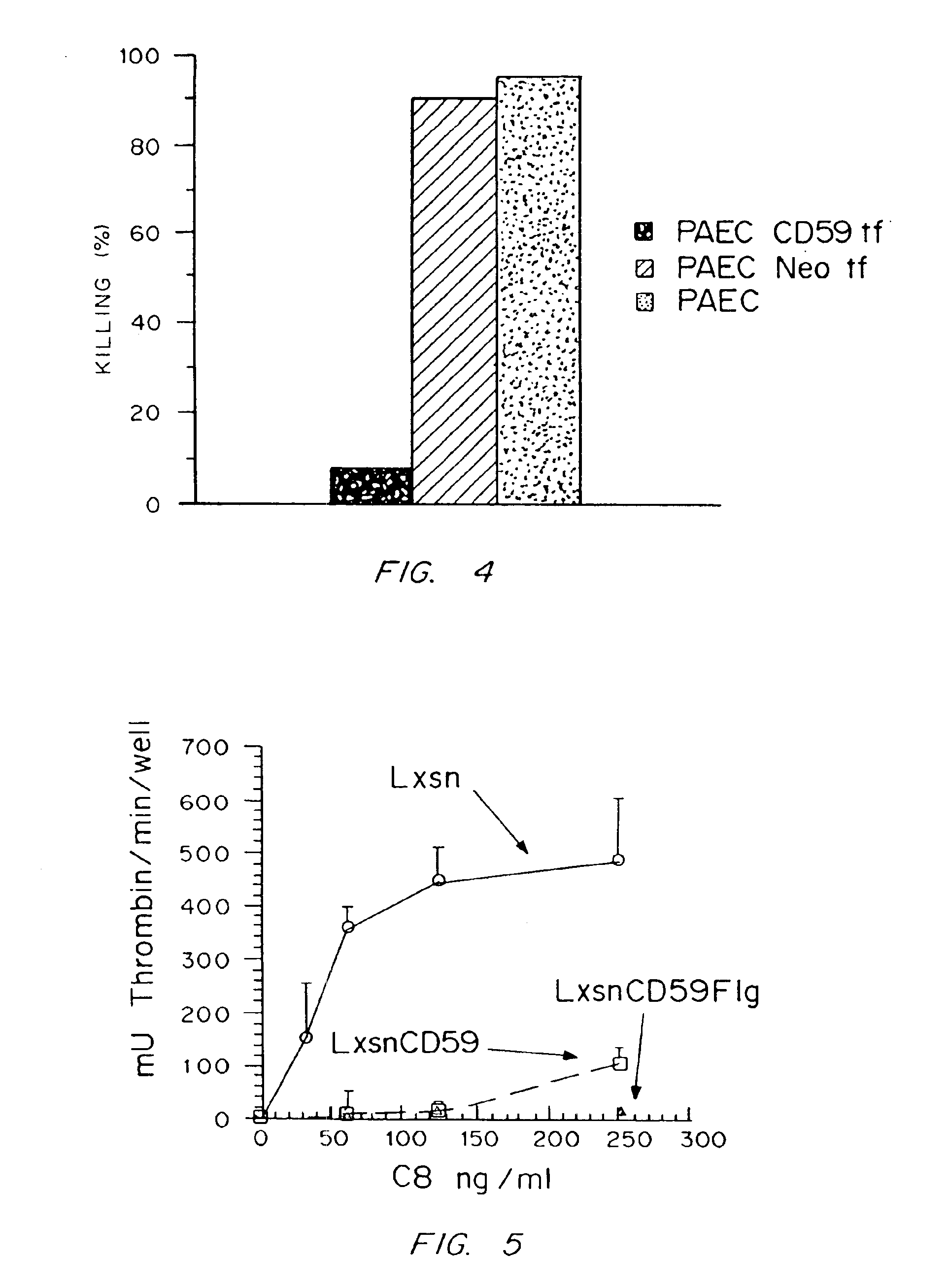
[0172]This example demonstrates that when the full-length CDNA encoding the human CD59 protein is stably incorporated into the genome of a porcine aortic endothelial cell (PAEC) and expressed on the cell surface, it protects these cells from complement-mediated attack as assayed by human complement-mediated cell lysis in vitro.

[0173]Cultures of PAEC were cultured in DMEM containing 10% fetal bovine serum (FBS), 5 mM Hepes, 2 mM L-glutamine, and 1% each of penicillin and streptomycin (P / S). Prior to retroviral infection, the cells were grown to 50% confluence. Subconfluent PAEC were infected by using the amphotropic helper-free retroviral vector pRNSRalphaCD59+. The structure of this retroviral construct is shown in FIG. 1. As controls, PAEC were also infected with a control retroviral vector containing the drug selection marker gene neomycin or were uninfected. The ampho...

example 2

Stable Expression of hCD59 in Mouse Cells and which Protects the Cells from Complement Mediated Lysis.

[0180]Mouse Balb / 3T3 cells were t-ransfected with the pC8-hCD59-103 plasmid described in FIG. 6. The transfection was performed using the calcium phosphate precipitation method with 10 μg of plasmid pC8-hCD59-103 applied to approximately 106 cells. Stable transfectants were selected by cotransfecting the Balb / 3T3 cells with a neomycin resistance plasmid (pSV2-neo; Yale University, New Haven, CT; 1 μg per 106 cells) and then selecting stably expressing cell clones in the presence of qeniticin (G418; 500 μg / ml). Balb / 3T3 cells transformed with just the neomycin resistance plasmid and not the pC8-hCD59-103 plasmid were used as controls.

[0181]Stable transfectants were assessed for hCD59 expression by flow cytometry using 10 μ / ml anti-CD59 monoclonal antibody MEM-43 (Accurate Chemical and Scientific Corp., Westbury, N.Y.) or 20 μg / ml of an anti-CD59 polyclonal serum (Southeastern Wiscons...

example 3

Generation of Transgenic Mice Expressing hCD59.

[0185]In order to achieve transgenic expression of hCD59, the CMV-hCD59-SV40 transcriptional unit was removed from pCB-hCD59-103 using the restriction enzymes SpeI and ScaI. This restriction digest selectively extracts the transcription unit from the overall vector and permits purification of only the essential elements-required for expression.

[0186]The 2300 bp restriction fragment resulting from the digest was gel isolated, extensively purified through an ELUTIP column (Schleicher & Schuell, Keene, N.H.), dialyzed against pyrogen free injection buffer (10 mM Tris, pH7.4+0.1 lmM EDTA in pyrogen free water), and used for embryo injection to generate transgenic mice in accordance with the methods of Hogan et al. 1986 and Brinster et al., 1985.

[0187]50 founder putative positive offspring were tested by harvesting genomic DNA from the tail of each of these animals. 5 micrograms of this genomic DNA from each of the 50 animals was transferred...


PUM

PropertyMeasurementUnit
cell doubling timeaaaaaaaaaa
pHaaaaaaaaaa
pHaaaaaaaaaa

Abstract

Genetically engineered cells are provided which can serve as universal donor cells in such applications as reconstruction of vascular linings or the administration of therapeutic agents. The cells include a coding region which provides protection against complement-based lysis, i.e., hyperacute rejection. In addition, the cell's natural genome is changed so that functional proteins encoded by either the class II or both the class I and the class II major histocompatibility complex genes do not appear on the cell's surface. In this way, attack by T-cells is avoided. Optionally, the cells can include a self-destruction mechanism so that they can be removed from the host when no longer needed.

Description

[0001]This is a continuation of U.S. Ser. No. 08 / 483,433 filed Jun. 7, 1995, now U.S. Pat. No. 6,100,443, which is a divisional of U.S. Ser. No. 08 / 087,007 filed Jul. 1, 1993, now U.S. Pat. No. 5,705,732, which is a continuation-in-part of U.S. Ser. No. 07 / 906,394 filed Jun. 29, 1992, now abandoned entitled “Universal Donor Cells” by Peter J. Sims, Alfred L.m. Bothwell, Eileen A. Elliot, Richard A. Flavell, Joseph Madri, Scott Rollins, Leonard Bell and Stephen Squinto.[0002]The U.S. Government has rights in this invention by virtue of Grants Nos. GM40924, 5 K11 HCO 2351, RO1 HL 28373, and 9T32 DK07556, awarded by the Department of Health and Human Services.BACKGROUND OF THE INVENTION[0003]This invention relates to genetically engineered endothelial cells and, in particular, to endothelial cells which have been modified to resist lysis and activation by complement and evade the host's immune mechanisms for removing foreign cells, when inserted into a non-autologous host.[0004]Endothe...

Claims


Application Information

Patent Timeline
Patent Type & Authority Patents(United States)
IPC IPC(8): A61L27/50A61L27/00C07K14/435C07K14/74C07K14/705C12N15/85A61K35/12A61K38/00A61K48/00A01K67/027
CPCA61L27/507C07K14/705C07K14/70539C07K14/70596C12N15/8509A01K67/0271A01K67/0275A01K67/0276A01K67/0278A01K2207/15A01K2217/00A01K2217/05A01K2217/075A01K2227/105A01K2267/025A01K2267/03A61K38/00A61K48/00A61K2035/122C12N2517/02
Inventor SIMS, PETER J.BOTHWELL, ALFRED L. M.ELLIOT, EILEEN A.FLAVELL, RICHARD A.MADRI, JOSEPHROLLINS, SCOTTBELL, LEONARDSQUINTO, STEPHEN
Owner OKLAHOMA MEDICAL RES FOUND
Why Eureka
Try Eureka