Supersensitive nuclear magnetic resonance micro imaging apparatus

a nuclear magnetic resonance and micro-imager technology, applied in the direction of reradiation, measurement using nmr, instruments, etc., can solve the problems of insufficient understanding of the correlation between the function and the structure of a protein molecule at the biocellular level, the growth of a high-quality protein crystal that can be analyzed sufficiently, and the need for several months to several years to grow. , to achieve the effect of extreme performance, increased throughput and measurement sensitivity

Inactive Publication Date: 2005-08-30
OKADA MICHIYA +4
View PDF8 Cites 10 Cited by
  • Summary
  • Abstract
  • Description
  • Claims
  • Application Information

AI Technical Summary

Benefits of technology

[0019]In the case where analysis using on-site observation of the metabolic reaction in the cells or imaging of a protein information network, which is considered to be increasingly required in the future, is performed effectively and accurately, experientially, it is desirable to be able to effect a measurement at 600 to 900 MHz, with 14 to 21 T at the central magnetic field, and with an appropriate amount of sample, and to obtain a sensitivity of measurement higher than the status quo and simultaneously to increase the throughput.
[0020]The object of the present invention is to provide a novel nuclear magnetic resonance analyzing apparatus, in which a sampling chamber of 1 to 30 mm in diameter that can accommodate the needs of biological study, is used, and in which the sensitivity for measuring the NMR signal is increased to a value in the order of at least three times the conventional value at least at 600 MHz (14.1T), and in which a time-based stability and spatial uniformity of the superconducting magnet required for imaging analysis in the cell can be provided.
[0021]In the construction of the present invention, the operating temperature of the system is not limited to 4.2 K. It is also possible to achieve extreme performance by the application of the present invention, and, depending on the applications, operation at 1.8K at 21.1T or 900 MHz, which used to be the limit of the magnetic field, is also possible. In such a case, improvement of the sensitivity to a value three times that currently available is possible, and thus breakthrough of the limit of detection sensitivity by the magnetic field strength, which used to be impossible, can be achieved for the first time.
[0022]The inventors have investigated the problems common to the present high-resolution nuclear magnetic resonan

Problems solved by technology

According to this publication, however, when the sensitivity is important, as in the case of measuring a minute amount of protein dissolved in an aqueous solution, it is impossible to wind a solenoid coil around a sample tube placed with a vertical orientation with respect to the magnetic field in the currently available superconducting magnet structure as a matter of fact, and thus such a coil is not generally used.
However, a method of improving the detection sensitivity required for high resolution imaging and a technical application for obtaining a uniformity of the magnetic fields or time-based stability of the magnetic field are not disclosed.
Remarkable progress has been made in recent years in the life sciences, but an understanding of the correlation between the function and the structure of a protein molecule at the biocellular level is not sufficient.
In addition, it is quite important for the progress of the life sciences to obtain a high-quality monocrystal of protein, but the growth of a high-quality protein crystal that can be analyzed sufficiently is still difficult, and the growth thereof requires several months to several years.
However, the spatial resolution of the current nuclear magnetic resonance analyzing apparatus is generally 0.2 mm, and thus a minute area in the order of 1 to 10 micron, which represents the sizes of cells, cannot be imaged.
When the magnetic fields are not uniform, problems, such as difficulty in identification of images, may occur.
Therefore, the performance of the NMR apparatus that has been generally

Method used


Image

  • Supersensitive nuclear magnetic resonance micro imaging apparatus
  • Supersensitive nuclear magnetic resonance micro imaging apparatus
  • Supersensitive nuclear magnetic resonance micro imaging apparatus

Examples


embodiment 1

(Embodiment 1)

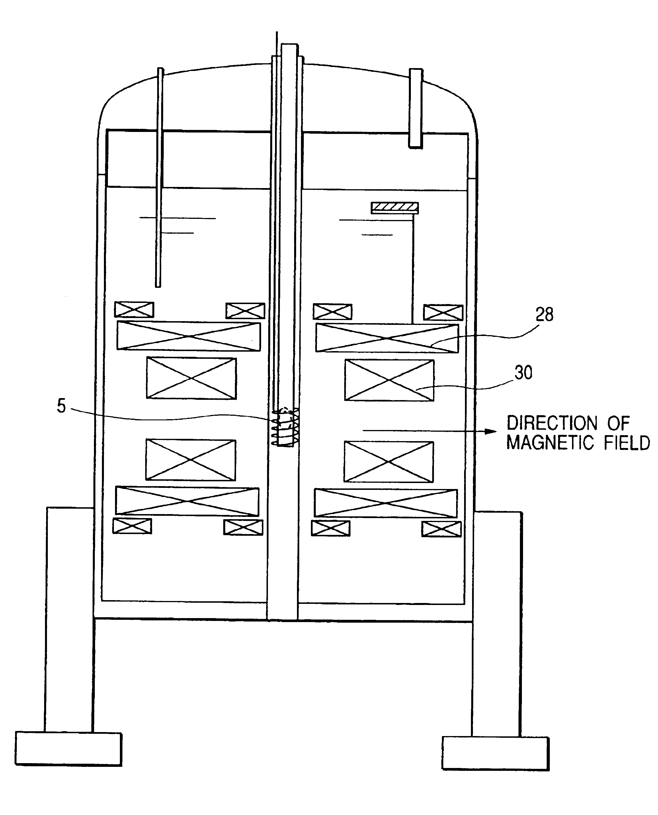
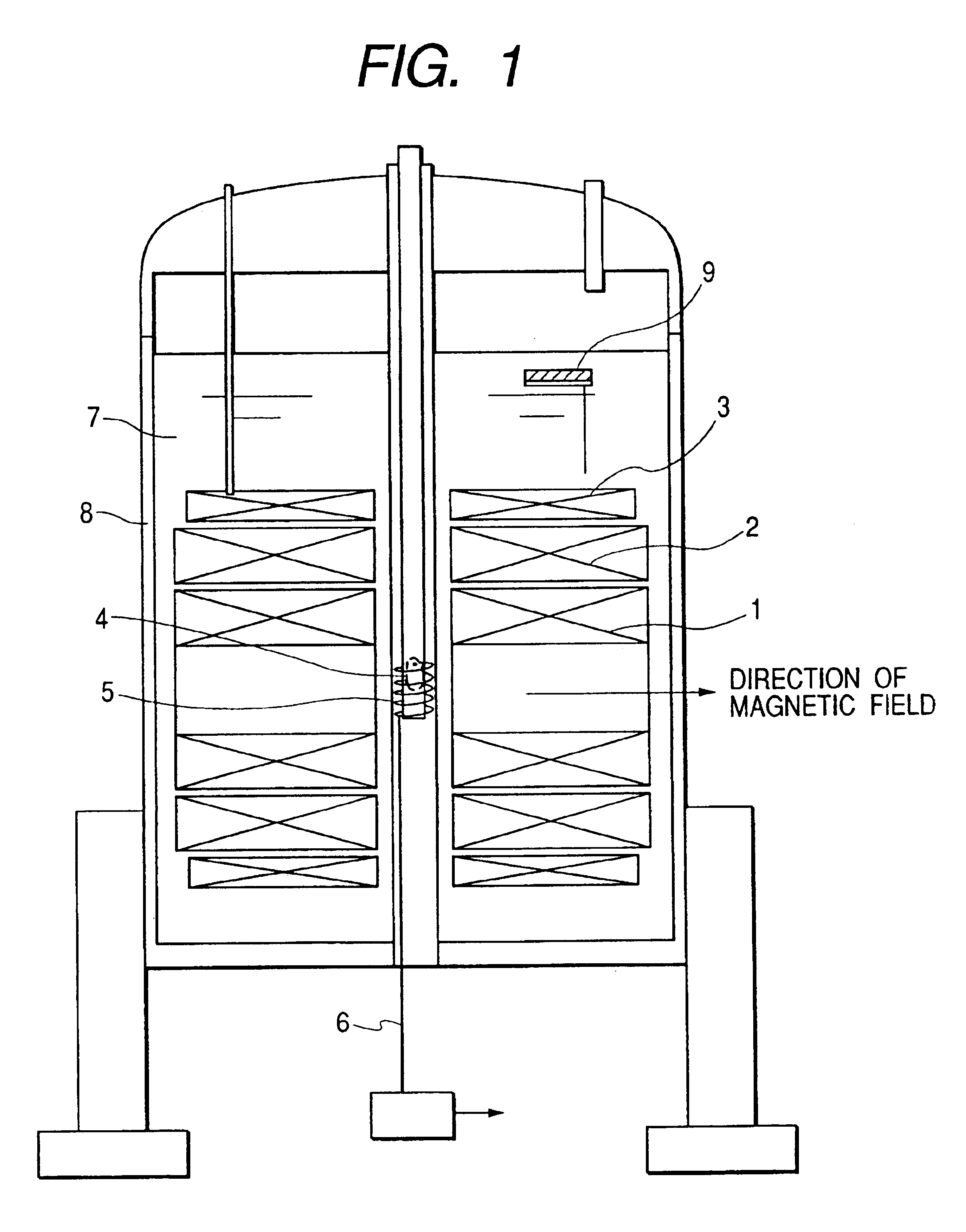
[0049]The first embodiment of the present invention is shown in FIG. 1. In the superconducting magnets 1, 2, 3 the coils are formed in such a manner that the inner side, that is the side closer to the sample, is formed of a material having a higher superconducting critical magnetic field. or example, the superconducting magnet 1 is formed of Nb3Al, the superconducting magnet 2 is formed of Nb3Sn, and the superconducting magnet 3 is formed of NbTi; and, they can be optically combined to obtain desired values of coil-generated magnetic field and of uniformity as needed. For example, a Bi-type such as Bl2Sr2CaCu2O9 and the like, or superconducting material, such as, Y1Ba5Cu3O7 and the like, may be used, or MgB2 and the like may also be used. The direction of generation of the magnetic fields of the supercoducting magnet 1, 2, 3 constructed of a combination thereof is horizontal.

[0050]In FIG. 1, the biosample 4, such as small animals, cells, and organic tissues, is inserte...

embodiment 2

(Embodiment 2)

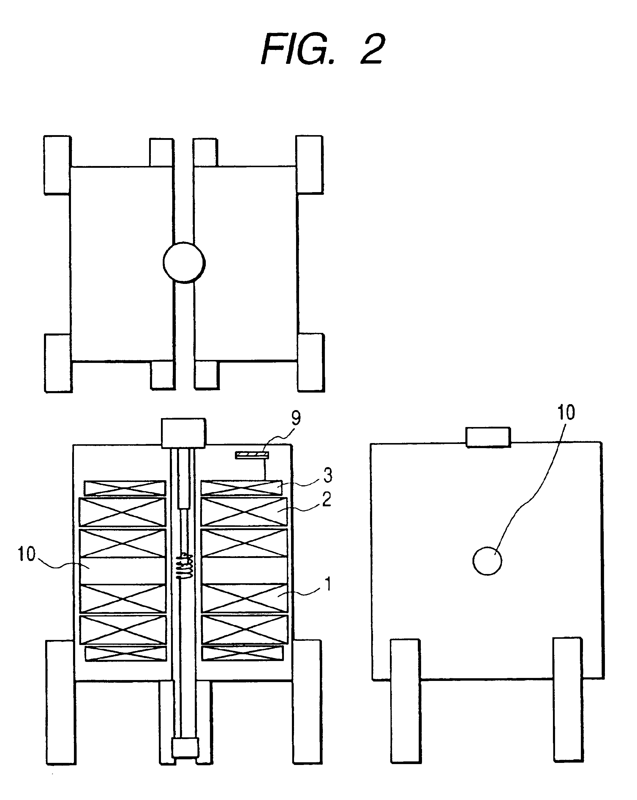
[0059]The second embodiment of the present invention is shown in FIG. 2. In this embodiment, the construction is generally identical to the first embodiment, but the low temperature container is divided by the left and right superconducting magnets to provide an openness to the available space for the user. In other words, since there is an open space 10 around the sample chamber, unlike the hermetical sample space in the previously known construction, the dynamic behavior of the liming organisms, such as photosynthesis, can be measured, while performing light irradiation or laser beam irradiation on the sample 4 easily. Since the dynamic NMR signal can be observed in this manner, for example, the signal transmission or reaction of photosynthesis of the protein can be inspected.

[0060]When performing such special experiments, liquid helium 7 is pumped to cool down and operate the apparatus at 1.8 K, so that the superconducting magnet 1, 2, 3 can be operated approximatel...

embodiment 3

(Embodiment 3)

[0061]A third embodiment of the present invention is shown in FIG. 3 in this embodiment, the biosample 4 is inserted into the apparatus from the top, and the measurement probe is inserted from the side. In this arrangement, the frame 11, that is provided with an anti-vibration device, can be lowered; and, thus the height of the apparatus cars be lowered, whereby the operability is improved, and vibrations propagated from the floor to the apparatus can be reduced. Therefore, an economically effective system, which is superior in installability and maintainability, can be provided.


PUM


Abstract

A supersensitive nuclear magnetic resonance imaging apparatus includes superconducting magnet, a gradient magnetic field coil, a high frequency emitting coil, and a receiving coil, wherein a biosample, including at least one of cells, organic tissues, and laboratory small animals, is inserted in a sample chamber of generally 1 to 30 mm in diameter. The superconducting magnet is formed of laterally divided split magnets, and the direction of the magnetic field generated by the magnet is generally horizontal. The receiving coil is in the form of a solenoid coil, and the biosample is inserted from a direction orthogonal to the direction of the magnetic field in a generally vertical direction. The spatial resolution in imaging of the biosample is not more than one-tenth of a cell that forms the biosample.

Description

CROSS-REFERENCE TO RELATED APPLICATION[0001]This is a continuation of U.S. application Ser. No. 10 / 326,085, filed Dec. 23, 2002, the subject matter of which is incorporated by reference herein.BACKGROUND OF THE INVENTION[0002]The present invention relates to a supersensitive nuclear magnetic resonance imaging apparatus; and, more particularly, the invention relates to an apparatus for effecting a high resolution imaging of biosamples, such as cells, organic tissues, laboratory small animals, or the like, and to growth of a high-grade protein crystal using high resolution imaging, on-site observation of the process of growth, and a method of growth.[0003]In recent years, rapid advances have been made in an imaging method utilizing nuclear magnetic resonance (NMR) imaging. If an analysis of the metabolism of cells or a protein information network in living organisms, such as human bodies, small animals, or cellular structures, can be made possible in the future by combining the NMR me...

Claims


Application Information

Patent Timeline
IPC IPC(8): G01R33/56G01R33/54G01R33/465G01R33/30G01R33/34G01R33/3815G01R33/48G01V3/00
CPCG01R33/3815G01R33/5604G01R33/465
Inventor OKADA, MICHIYAWAKUDA, TSUYOSHIKAKUGAWA, SHIGERUMORITA, HIROSHIAIHARA, KATSUZOU
Owner OKADA MICHIYA
Why Eureka
Try Eureka