
Vivo monitoring method of transgenic plants and system using the same

a monitoring method and plant technology, applied in the field of transgenic plant monitoring methods and systems, can solve the problems of low light intensity of luc, difficult to directly observe the exogenous fluorescence of proteins introduced into plant tissues under fluorescent optical microscopes, and inconvenient direct visual selection of transgenic plants

Inactive Publication Date: 2005-09-20
DONGBU HANNONG CHEMICAL CO +1
View PDF7 Cites 79 Cited by
  • Summary
  • Abstract
  • Description
  • Claims
  • Application Information

AI Technical Summary

Benefits of technology

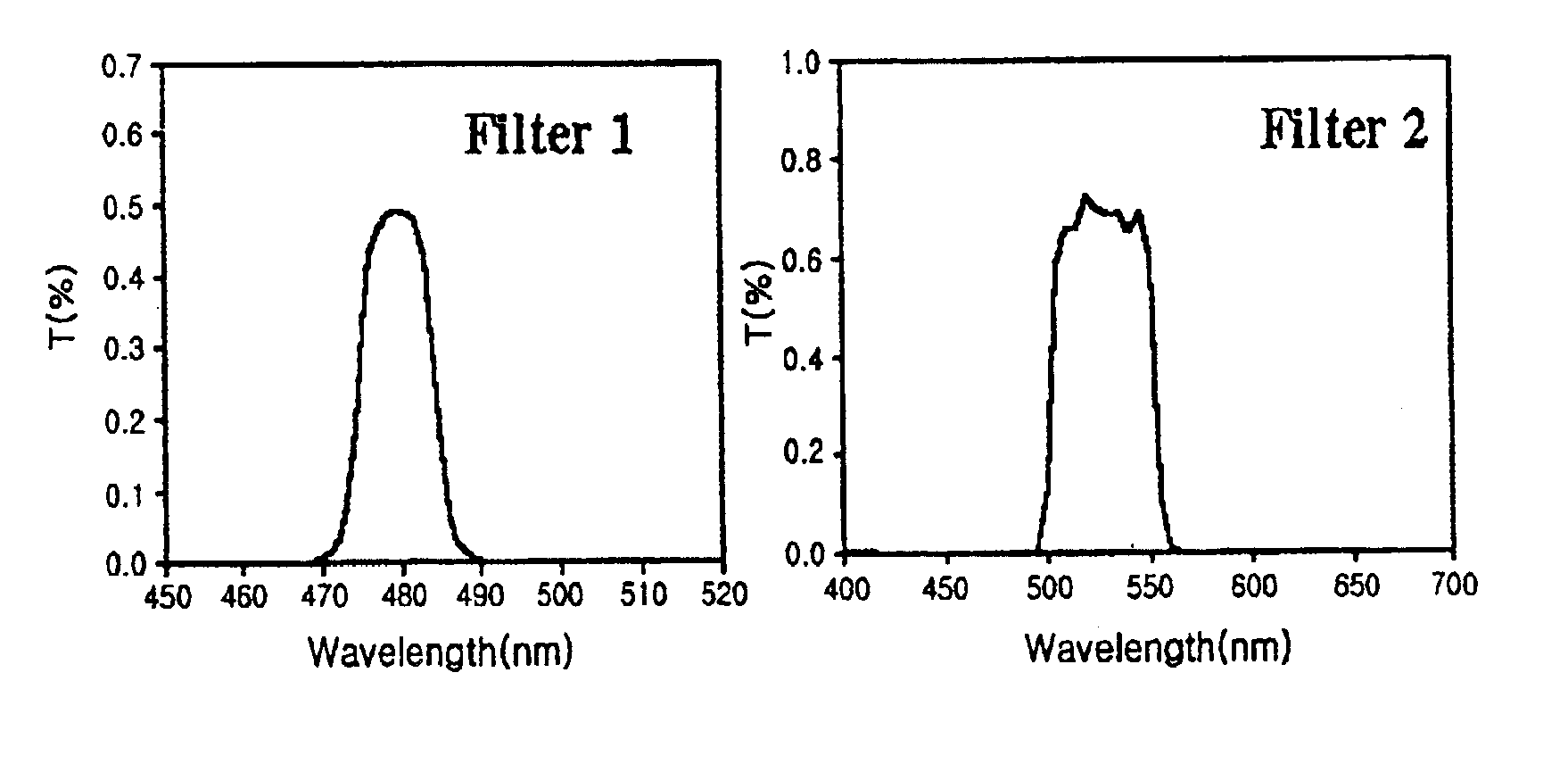
[0005]Leading to the present invention, the intensive and thorough research on the identification of transgenic plants, conducted by the present inventors, resulted in the finding that, when a filtered light beam which can excite GFP is projected to a plant sample of interest, light is emitted from the plant sample and, if filtered through a green bandpass filter, can be analyzed for the expression of GFP by use of a CCD (charge-coupled device) camera imaging system.

Problems solved by technology

However, histochemical GUC analysis is not suitable for the direct visual selection of transgenic plants per se because it requires the destruction of plant tissues.
In addition, LUC has the disadvantage of emitting a low intensity of light (Ow D W et al., Science 234:856-859).
Because of plant tissues' being composed of highly reflective cell walls and aqueous cytoplasm containing various autofluorescent and light-scattering materials, it is difficult to directly observe the exogenous fluorescence of proteins introduced into plant tissues under a fluorescent optical microscope (Haseloff J. et al., 1998, Green fluorescent protein: Properties, applications, and protocols.

Method used


Image

  • Vivo monitoring method of transgenic plants and system using the same
  • Vivo monitoring method of transgenic plants and system using the same
  • Vivo monitoring method of transgenic plants and system using the same

Examples


example 1

Construction of Vector

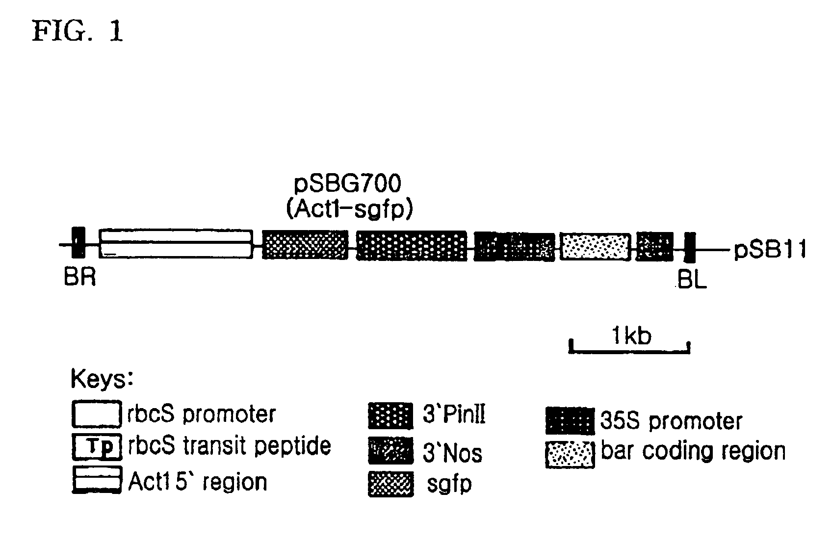
[0029]After being digested with BamHI and NcoI, a rice-derived Act1 promoter (McElroy D. et al., 1991, Mol. Gen. Genet. 231:150-160 was inserted into a BamHI / NcoI-linearized pBluescript plasmid containing an sgfp gene (Kohler R H. et al., 1997, Plant J. 11:613-621). Treatment of the resulting recombinant plasmid with BamHI and NotI extracted an Act1 promoter-sgfp fragment. This DNA fragment was ligated to a BamHI / NotI-linearized pSB105 that contained potato proteinase inhibitor II terminator / 35S promoter / bar / nopaline synthase terminator to construct a recombinant plasmid, named pSBG700, as illustrated in FIG. 1. Using the tripatental mating method disclosed in Komari T., et al., 1996, Plant J. 19:165-174), Agrobacterium tumefacience LBA4404 was transformed with the plasmid pSBG700.

example 2

Transformation of Rice

[0030]A wild-type rice seed (Oryza sativa cv. Nakdong) and a rice seed transformed with an agfp gene (Jang et al., 1999, Molecular breeding 5:453-461) were treated with 70% ethanol for 1 hour and then with 10% sodium hypochlorite for 15 min. After being washed five times with sterile water, the seeds were sowed in test tubes containing MS salt (Murashige T., Skoog F., 1962, Physiol. Plant, 15:473-497), sucrose 30 g / l, and bactoagar 8 g / l. Subsequently, the test tubes were incubated in a growth chamber which was adjusted to a temperature of 25° C. with a PFD maintained at 100 μmol m−2s−1. Under light-illuminated and shielded conditions, the seeds were let to grow for 16 hours and 8 hours, respectively. The seeds were germinated to white plants in the test tubes in the dark. In petri dishes containing MS salt, glucose 30 g / l, 2,4-D 2 mg / l, and bactoagar 8 g / l, calluses were induced from the white sprouts and grown at 25° C. in the growth chamber under the control...

example 3

GFP Fluorometry

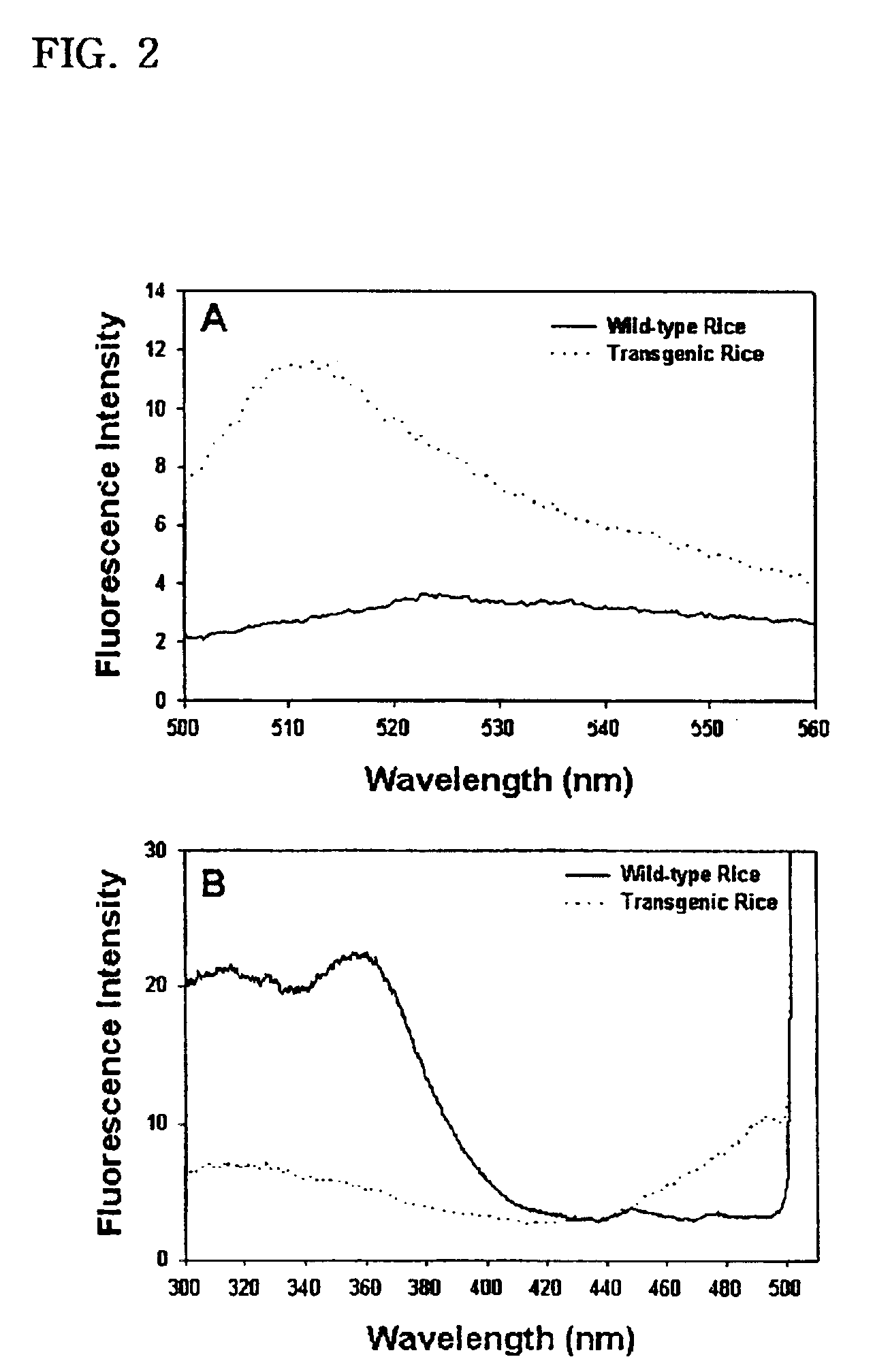
[0032]To determine the excitation and emission spectra of the GFP that the transgenic plants produced, water-soluble proteins were extracted from leaves of the transgenic rice which had been grown for three months.

[0033]To this end, first, sliced leaves were homogenized in an extraction buffer (20 mM Tris-HCl, pH 8.0, 10 mM EDTA, 30 mM NaCl, 2 mM phenylmethanesulfonyl fluoride). After the centrifugation of the homogenate at 12,000×g for 10 min, a portion of the supernatant thus obtained was added to the extraction buffer to make a final volume of 1 ml. Using an assay kit (Bio-Rad) according to manufacturer's instruction, water-soluble proteins were quantitatively analyzed with bovine serum albumin serving as a control.

[0034]At room temperature, a quantitative measurement was made of the GFP fluorescence of cell extracts with the aid of an F-450 fluorometer (Hitachi, Japan) in 10 mm / 10 mm cuvettes. After passing through the excitation and emission monochromator used in...


PUM

PropertyMeasurementUnit
wavelengthaaaaaaaaaa
angleaaaaaaaaaa

Abstract

The present invention relates to a method for visualizing GFP expression in callus, various tissue and organ of the transgenic plants as image and system using the same. The said method needs no other additional genetic product, substrate or cofactor and can detect very simply and quickly GFP expression by using the said system of the present invention consisting of a CCD camera, a light source, band-pass filter and data processing computer, so it provides many advantages for selection of transgenic seeds, for studying of gene expression in the tissue or organ of plants, or for studying of specificity of each development step.

Description

TECHNICAL FIELD[0001]The present invention relates to a method and system for monitoring the transformation of plants. More particularly, the present invention relates to a fluorometry method and system for the visualization of the in vivo expression of a green fluoroscent protein gene tagged to an exogenous gene of interest, thereby enabling the plants to be determined as to whether they are transformed with the exogenous gene or not.BACKGROUND ART[0002]Reporter proteins are usually used as markers for visualizing in vivo gene expression and protein translocation in eucaryotic and prokaryotic organisms. The most widely used reporter proteins in plants may be exemplified by β-glucuronidase (GUS) and luciferase (LUC). Particularly, GUS is a prevalent visualization marker in the plant cell biology (Jefferson R A, et al., (1987), EMBO J., 6:3901-3907). However, histochemical GUC analysis is not suitable for the direct visual selection of transgenic plants per se because it requires the...

Claims


Application Information

Patent Timeline
Patent Type & Authority Patents(United States)
IPC IPC(8): G01N21/64G01N33/48
CPCG01N21/6428G01N33/48
Inventor KIM, JOO-KONCHUNG, BYOUNG-CHULLPARK, YONG-JOO
Owner DONGBU HANNONG CHEMICAL CO
Learn More