

Imaging of drug accumulation as a guide to antitumor therapy
a drug accumulation and imaging technology, applied in the field of radiolabeled antitumor drugs, can solve the problems of large variability in the efficacy of treatments, small tissue sample, and often extremely toxic cancer therapies for patients, and achieve the effect of efficient determination of potentially efficacious treatment plans
- Summary
- Abstract
- Description
- Claims
- Application Information
AI Technical Summary
Benefits of technology
Problems solved by technology
Method used
Image

Examples
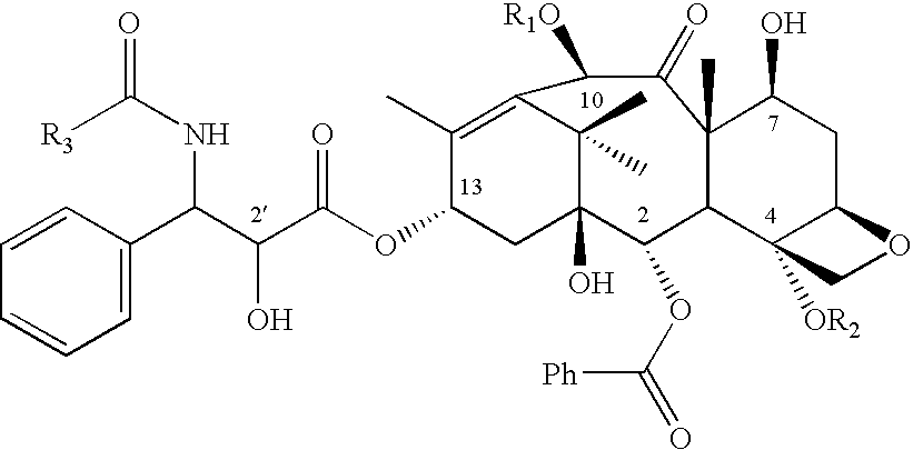
example 1
Synthesis of 11C-paclitaxel
[0122]10-Deacetylpaclitaxel (6 mg) (commercially available) in pyridine (0.5 ml) was reacted with chlorotriethylsilane (0.1 ml) for 1 hour at 60° C. to yield 7,2′-di-(triethylsilyl)--10-deacetylpaclitaxel. This molecule is the immediate precursor for radiolabeling; it is stable at room temperature and can be stored until needed.
[0123]To a solution of (50 μg, 60 nmol) of 7,2′-di-(triethylsilyl)-10-deacetylpaclitaxel, was added dimethylaminopyridine solution (90μl of 300 mg / ml in methylene chloride), and of tert-butyl diphenyl chlorosilane (20 μL). Acetyl chloride (10 μl of a 1:5000 solution in methylene chloride, 25 nmol) was then added and the mixture heated to 105° C. for 10 minutes. The silyl groups were removed within 2 minutes by adding methylene chloride (400 μl) and tetrabutylammonium fluoride solution (50 μl of 1 M solution in tetrahydrofuran). Verification of the identify of the product via HPLC and mass spectrometry was obtained by comparison with...
example 2
Alternative Synthesis of 11C-paclitaxel
[0126]Benzoyl chloride (8 μl of 1:1000 dilution in acetonitrile) is added to a solution of paclitaxel primary amine (50 μg) (commercially-available) in acetonitrile (200 μl). After 2 minutes at room temperature, 60% recovery of product was obtained.
[0127]Conversion of this procedure to radio-synthesis requires preparation of 11C-benzoyl chloride, which is known in the art by following typical Gringard reaction schemes. Mathews W B, Burns H D, Danals R F, Rabert H T, Naylor E M. Carbon-11 labeling of a potent, nonpeptide, AT1-selective angiotensin-11 receptor antagonist, MK-996. J. Labelled Compounds and Radiopharmaceuticals 1995; XXXVI:729–37. For 11C-paclitaxel, purification may be accomplished by HPLC or by adding acidic water and methylene chloride.
example 3
Synthesis of 11C-docetaxel
[0128]Preparation of Docetaxel Primary Amine
[0129]The starting material for the preparation of 11C-docetaxel is docetaxel primary amine, which is not available commercially. The free amine is prepared by removal of the tert-butyl carbonyl group from docetaxel using formic acid or other methods known in the art.
[0130]Docetaxel was purified from its commercial form (TAXOTERE® for Injection RhonePoulencRorer) by silica gel chromatography. Docetaxel (4.5 mg) was dissolved in EtOH (1 ml). After addition of formic acid (1 ml) the mixture was stirred at room temperature for 10 days. Overall conversion to docetaxel primary amine was 48%. Water (2 ml) and methylene chloride (2 ml) were added to the reaction solution and mixed. The aqueous phase containing docetaxel primary amine was separated from the organic phase containing residual docetaxel. The aqueous phase was neutralized with saturated sodium bicarbonate in water. Methylene chloride (12 ml) was added to the ...
PUM
| Property | Measurement | Unit |
|---|---|---|
| half life | aaaaa | aaaaa |
| PET | aaaaa | aaaaa |
| resistance | aaaaa | aaaaa |
Abstract
Description
Claims
Application Information