Intrinsically fluorescent, self-multimerizing MHC fusion proteins and complexes thereof

a fusion protein and self-multimerizing technology, applied in the field of intra-fluorescence, self-multi-merizing mhc fusion proteins and to complexes thereof, can solve the problems of affecting the yield and utility of the resulting tetramer complex, and reducing the yield of tetramers, so as to achieve sufficient avidity for t lymphocytes and stable binding to surfa

Inactive Publication Date: 2007-04-10
BECTON DICKINSON & CO
View PDF7 Cites 16 Cited by
  • Summary
  • Abstract
  • Description
  • Claims
  • Application Information

AI Technical Summary

Benefits of technology

[0034]It is an object of the present invention to provide reagents that can be used to label T lymphocytes based upon their antigen (and MHC) specificity, that have sufficient avidity for T lymphocytes to permit stable binding to surface TCR without requiring biotinylation and use of an extrinsic multimerizing moiety, and that can be rendered fluorescent without requiring use of secondary antibodies or direct chemical conjugation to fluorophore. It is a further object of the invention to provide methods for labeling, detecting, enumerating, enriching, isolating, depleting, and activating antigen-specific T lymphocytes using such reagents.

Problems solved by technology

Although MHC tetramers have proven tremendously useful, certain aspects of their synthesis present problems.
Inefficiency or imprecision at either or both of these steps affects yield and utility of the resulting tetrameric complex.
For example, inefficient biotinylation of the MHC chain—i.e., failure to incorporate at least one biotin per MHC molecule—will lead to decreased yield of tetramers.
Inefficient removal of unconjugated biotin from the reaction products, with carry-over of unconjugated biotin molecules into the multimerization reaction, can saturate avidin molecules and also decrease tetramer yield.
Early attempts at multimerization using chemical biotinylation failed, possibly because multiple and random chemical modification of MHC molecules led to randomized, and often inactive, orientation of the MHC / peptide moieties in the multimerized complex.
Higher order multimers can also produce a more intense fluorescent signal, further leading to confounding variations in staining intensity.
Conversely, carry-over of unconjugated biotin molecules into the multimerization reaction can lead to partial saturation of avidin / streptavidin moieties with unconjugated biotin, leading to formation of multimeric complexes having fewer than four MHC / peptide subunits.
The requirement for a separate fluorophore conjugation step also presents opportunities for yield loss and for unintended variation in either avidity for TCR or fluorescence intensity, or both.
The self-dimerizing chimeras obviate the biotinylation step and use of an extrinsic multimerizing moiety required by MHC chimeras, with their attendant problems.
However, the dimeric chimeras are not without problems of their own.
First, the dimeric valency can cause lower avidity for TCR than would be obtained using tetramers, reducing or abrogating the ability to label low affinity TCRs.
Additionally, each of the strategies for rendering the dimeric chimera fluorescently detectable presents difficulties.
For example, use of secondary antibodies for visualization can make simultaneous detection of multiple antigens more difficult; this can be a significant problem when, as is often the case, the population of antigen-specific T cells is small, obligating use of multiple parameters in order accurately to detect and enumerate the cells.
Direct chemical conjugation of fluorophore to the chimera, an alternative to use of secondary antibodies, can interfere with or even abrogate affinity interactions of the chimera with its cognate TCR.
Direct chemical conjugation can also lead to chimeric molecules having varying molar ratios of label, making flow cytometric or other fluorescence-based analyses more problematic.
First, the red fluorescence of DsRed is slow to mature, requiring days to ripen fully from green to red, both in vitro and in vivo.
Although oligomerization may be irrelevant to use of DsRed as a simple reporter of gene expression, it will present serious problems in most potential applications where DsRed would be fused to a host protein to report on the trafficking or interactions of the latter.

Method used


Image

  • Intrinsically fluorescent, self-multimerizing MHC fusion proteins and complexes thereof
  • Intrinsically fluorescent, self-multimerizing MHC fusion proteins and complexes thereof
  • Intrinsically fluorescent, self-multimerizing MHC fusion proteins and complexes thereof

Examples


example 1

Construction of an Expression Vector for the Expression of a MHC-DsRed Fusion Protein

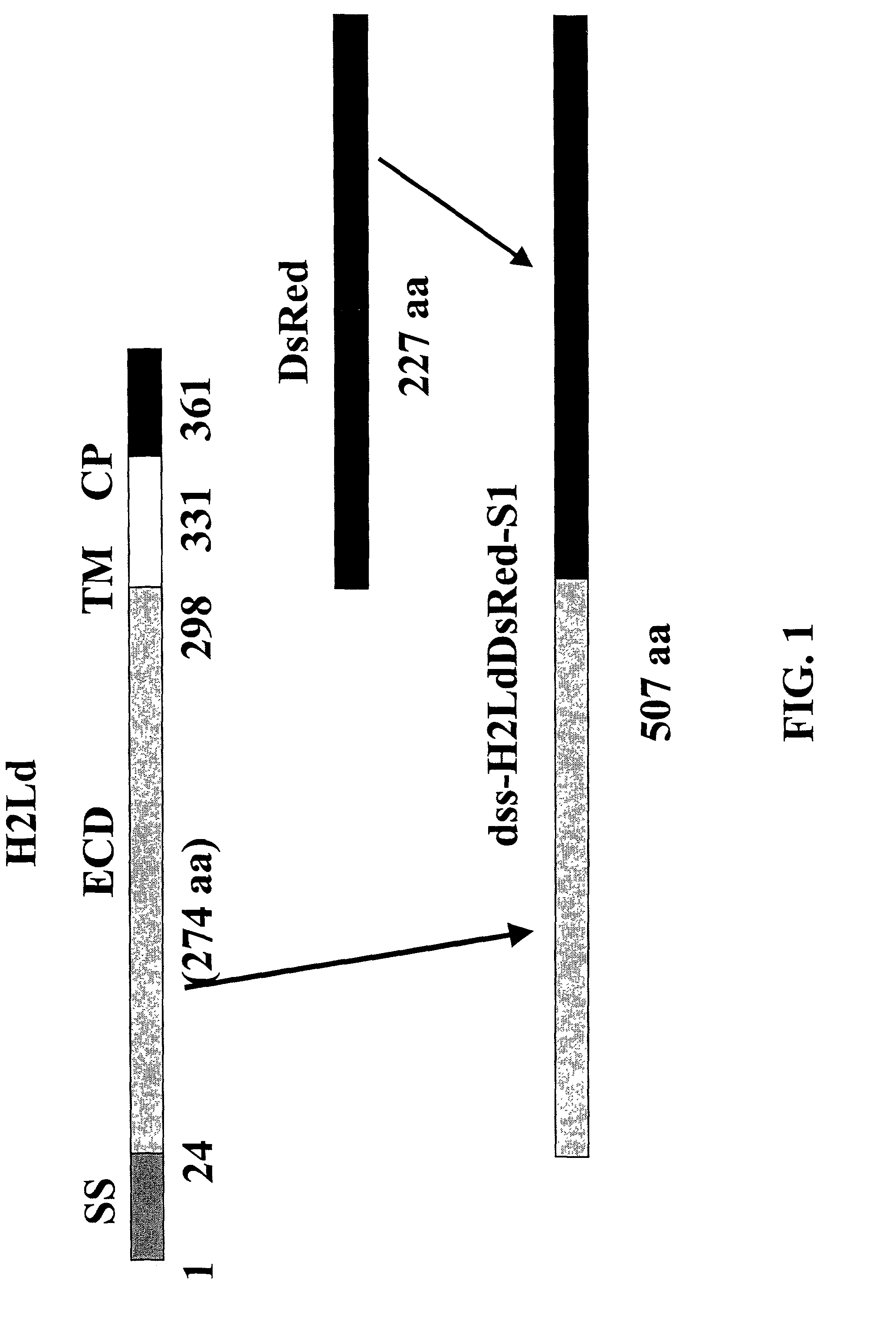
[0233]Using standard molecular biology techniques, a DNA sequence encoding the extracellular domain (ECD) of murine H2-Ld MHC class I protein is removed from the H2-Ld CDNA and ligated into the Multiple Cloning Site of the vector pDsRed2-N1 (BD Biosciences-Clontech Cat. #6973-1) digested with the enzymes HindIII and BamHI. This results in an in-frame fusion of the coding sequence of the ECD, oriented at the amino-terminal part of the fusion, with the coding sequence of the DsRed chromophore, oriented at the carboxy-terminal part of the fusion. Construction of the fusion construct is shown schematically in FIG. 1.

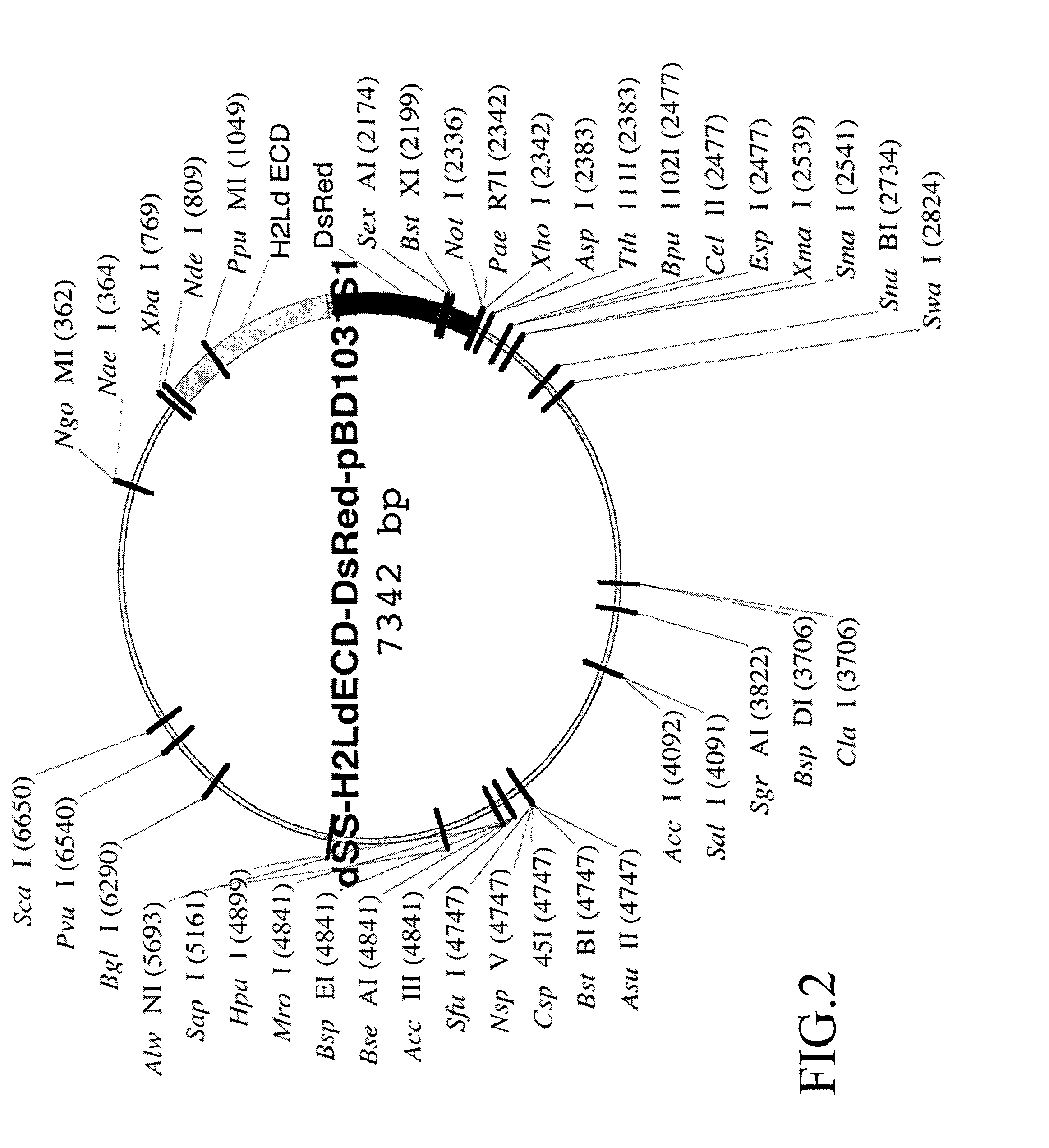
[0234]The H2Ld-DsRed2 fusion construct is then ligated into the NdeI and NotI restriction enzyme sites of pBD1031S1, a BD Biosciences-Pharmingen vector developed from PVL1393, Cat. #21201P. The pBD1031S1 vector is a dual expression vector containing a T7 RNA polymerase promoter for expressio...

example 2

Expression of the H2Ld-DsRed Fusion Protein in Bacteria

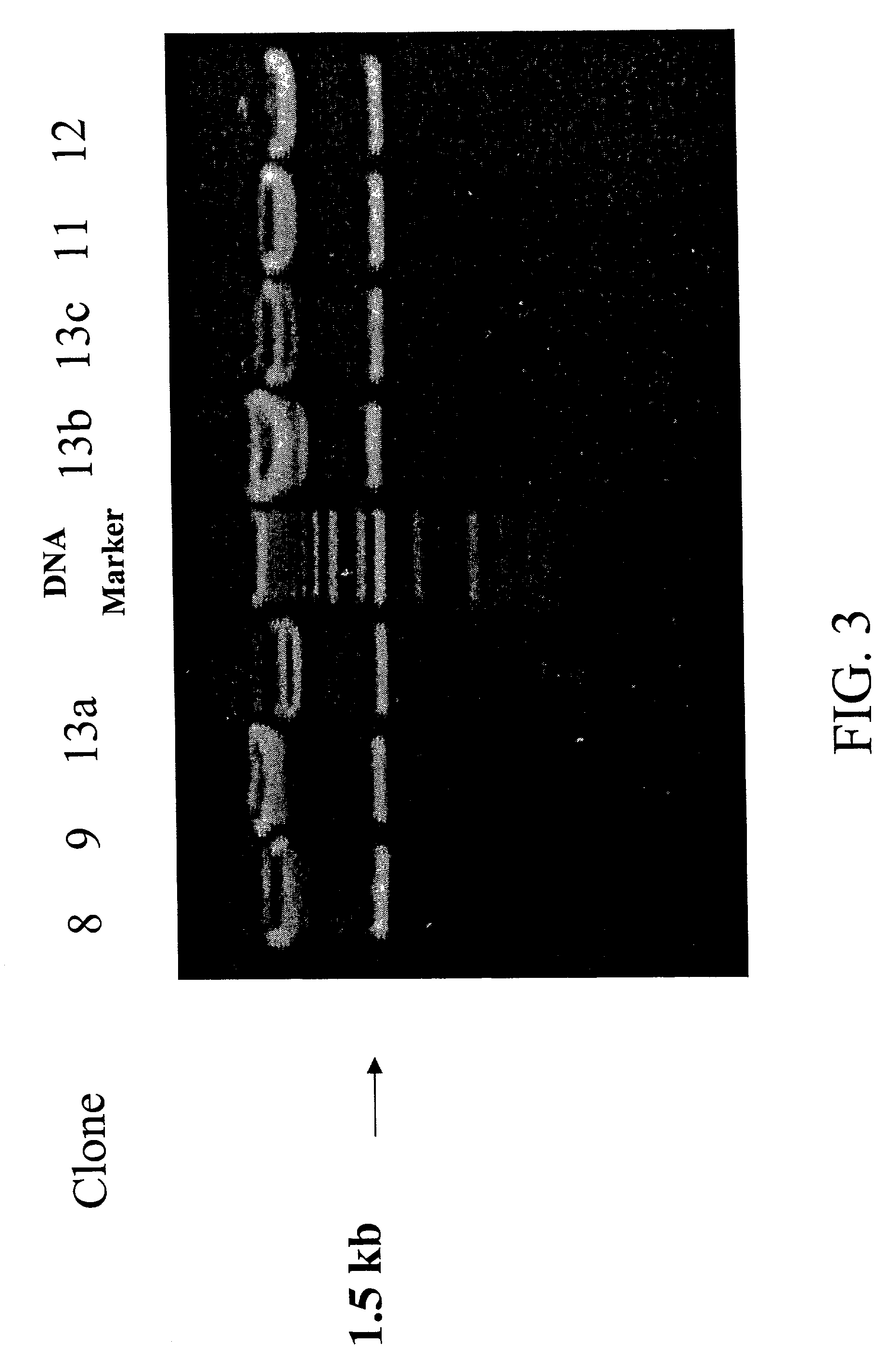
[0238]Using standard techniques, five clones of the H2Ld-DsRed fusion construct expression vector are used to transform competent E. coli bacteria containing an IPTG inducible T7 RNA polymerase. After growth overnight on ampicillin-LB agar plates, colonies of transformed cells are picked and expanded by growth overnight in LB media. The next day, the overnight culture is further grown at 37° C. in fresh LB media containing IPTG (final concentration 1 mM) for 2 hours.

[0239]Samples of bacteria from induced cultures are then withdrawn and lysed in an SDS-containing buffer, after which the constituent proteins are separated by SDS denaturing polyacrylamide gel electrophoresis (PAGE). After electrophoresis, gels are stained with Coomassie stain, after which the separated, stained proteins are illuminated by visible light and photographed with a video still camera for visual analysis.

[0240]As shown in FIG. 5, for each of five H2Ld-DsR...

example 3

Expression of the H2Ld-DsRed Fusion Protein in Insect Cells

[0242]The H2Ld-DsRed expression vector is cotransfected with BaculoGold™ DNA (BD-Pharmingen, Cat. No. 554739, formerly Cat. No. 21100D) into Sf9 and Tni insect cells using standard techniques discussed in the BD BaculoGold™ Linearized Baculovirus DNA technical data sheet, incorporated herein by reference in its entirety, and available from Pharmingen (BD Bioscience Pharmingen, San Diego, Calif. The expression vector serves as a transfer vector that complements a deletion in the BaculoGold™ virus DNA such that infectious virus is produced that contains within its genome the DNA encoding the H2Ld-DsRed fusion protein. Three days after transfection performed using standard techniques, medium from the transfected cell cultures is collected, cells are removed, and the supernatant is used in the first of three rounds of amplification to obtain a high titer stock solution of infectious baculovirus.

[0243]Using standard techniques, t...


PUM

PropertyMeasurementUnit
temperaturesaaaaaaaaaa
concentrationaaaaaaaaaa
temperaturesaaaaaaaaaa

Abstract

Presented are intrinsically fluorescent, self-multimerizing MHC fusion proteins, and complexes assembled therefrom that are capable of detectably labeling antigen-specific T lymphocytes. Also presented are methods for labeling antigen-specific T lymphocytes with the intrinsically fluorescent complexes of the present invention, and methods, particularly flow cytometric methods, for detecting, enumerating, enriching, and depleting antigen specific T lymphocytes so labeled.

Description

CROSS REFERENCE TO RELATED APPLICATION[0001]This application claims the benefit of the filing date of U.S. provisional application Ser. No. 60 / 261,448, filed Jan. 12, 2001, the disclosure of which is incorporated herein by reference in its entirety.FIELD OF THE INVENTION[0002]The present invention relates to intrinsically fluorescent, self-multimerizing MHC fusion proteins and to complexes thereof, and to methods for using such complexes for the labeling, detection, enumeration, enrichment, isolation, depletion and activation of antigen specific T lymphocytes, particularly by flow cytometry.BACKGROUND OF THE INVENTION[0003]Soluble tetramers of major histocompatibility complex (MHC) proteins, charged with specific peptide antigen and fluorescently labeled (“MHC tetramers”), have in the past five years proven extraordinarily useful in the detection, enumeration, characterization and purification of antigen-specific T lymphocytes. Applications of MHC class I tetramers have recently bee...

Claims


Application Information

Patent Timeline
Patent Type & Authority Patents(United States)
IPC IPC(8): C07K14/74C12N5/10G01N33/53G01N33/532C07K1/14C07K14/435C07K14/47C07K19/00C12N1/15C12N1/19C12N1/21C12N15/09G01N21/78G01N33/543
CPCC07K14/43595C07K14/70539C07K19/00C07K2319/00Y10S435/975
Inventor DAVIS, KENNETH A.WEI, BING-YUAN
Owner BECTON DICKINSON & CO
Why Eureka
Try Eureka