Nanoparticles for protein drug delivery

a technology of nanoparticles and proteins, applied in the field of nanoparticles, can solve the problems of severe restrictions, hydrophilic drug inability to easily diffuse across cells, and nanoparticles are not ideal carriers of hydrophilic molecules, and achieve the effect of reducing the inflammatory respons

Inactive Publication Date: 2011-04-05
NANOMEGA MEDICAL CORP
View PDF4 Cites 9 Cited by
  • Summary
  • Abstract
  • Description
  • Claims
  • Application Information

AI Technical Summary

Benefits of technology

[0032]Some aspects of the invention provide a method of reducing inflammatory response caused by tumor necrosis factor in an animal subject, the method comprising orally administering nanoparticles composed of a TNF inhibitor, chitosan, and a core substrate of poly(glutamic acid) or heparin.

Problems solved by technology

This is because hydrophilic drugs cannot easily diffuse across the cells through the lipid-bilayer cell membranes.
However, the transport of hydrophilic molecules via the paracellular pathway is, however, severely restricted by the presence of tight junctions that are located at the luminal aspect of adjacent epithelial cells (Annu. Rev. Nutr. 1995; 15:35-55).
However, these nanoparticles are not ideal carriers for hydrophilic drugs because of their hydrophobic property.
The absorption of protein drugs following oral administration is challenging due to their high molecular weight, hydrophilicity, and susceptibility to enzymatic inactivation.
However, the tight junction forms a barrier that limits the paracellular diffusion of hydrophilic molecules.
Most commercially available CSs have a quite large molecular weight (MW) and need to be dissolved in an acetic acid solution at a pH value of approximately 4.0 or lower, which is somewhat impractical.

Method used


Image

  • Nanoparticles for protein drug delivery
  • Nanoparticles for protein drug delivery
  • Nanoparticles for protein drug delivery

Examples


example no.1

Example No. 1

Materials and Methods of Nanoparticles Preparation

[0065]CS (MW ˜2.8×10⁵) with a degree of deacetylation of approximately 85% was acquired from Challenge Bioproducts Co. (Taichung, Taiwan). Acetic acid, cellulase (1.92 units / mg), fluorescein isothiocyanate (FITC), phosphate buffered saline (PBS), periodic acid, sodium acetate, formaldehyde, bismuth subnitrate, and Hanks' balanced salt solution (HBSS) were purchased from Sigma Chemical Co. (St. Louis, Mo.). Ethanol absolute anhydrous and potassium sodium tartrate were obtained from Merck (Darmstadt, Germany). Non-essential amino acid (NEAA) solution, fetal bovine serum (FBS), gentamicin and trypsin-EDTA were acquired from Gibco (Grand Island, N.Y.). Eagle's minimal essential medium (MEM) was purchased from Bio West (Nuaille, France). All other chemicals and reagents used were of analytical grade.

example no.2

Example No. 2

Depolymerization of CS by Enzymatic Hydrolysis

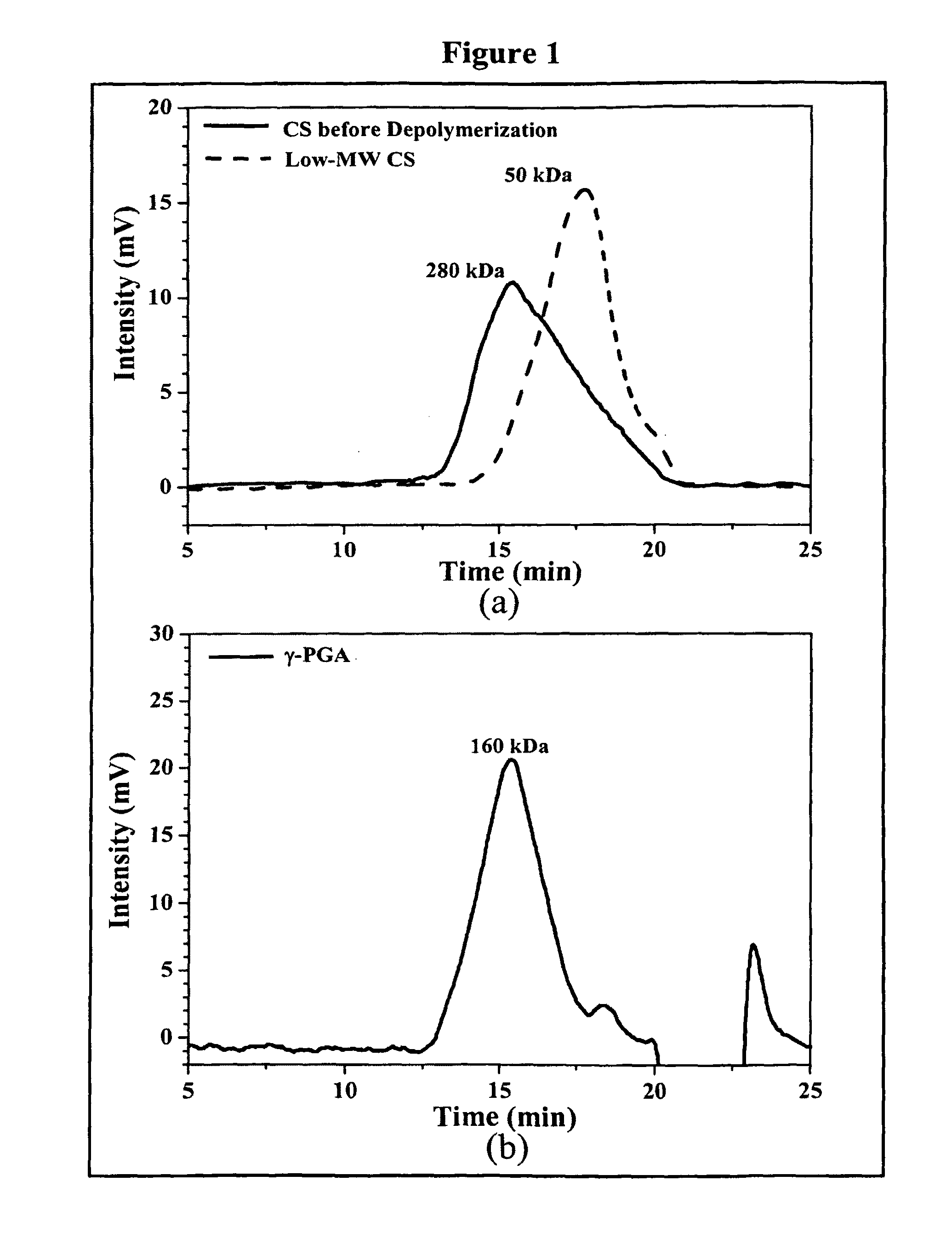
[0066]Regular CS was treated with enzyme (cellulase) to produce low-MW CS according to a method described by Qin et al. with some modifications (Food Chem. 2004; 84:107-115). A solution of CS (20 g / l) was prepared by dissolving CS in 2% acetic acid. Care was taken to ensure the total solubility of CS. Then, the CS solution was introduced into a vessel and adjusted to the desired pH 5.0 with 2N aqueous NaOH. Subsequently, cellulase (0.1 g) was added into the CS solution (100 ml) and continuously stirred at 37° C. for 12 hours. Afterward, the depolymerized CS was precipitated with aqueous NaOH at pH 7.0-7.2 and the precipitated CS was washed three times with deionized water. The resulting low-MW CS was lyophilized in a freeze dryer (Eyela Co. Ltd, Tokyo, Japan).

[0067]The average molecular weight of the depolymerized CS was determined by a gel permeation chromatography (GPC) system equipped with a series of PL aquagel-OH column...

example no.3

Example No. 3

Preparation of the CS-γ-PGA Nanoparticles

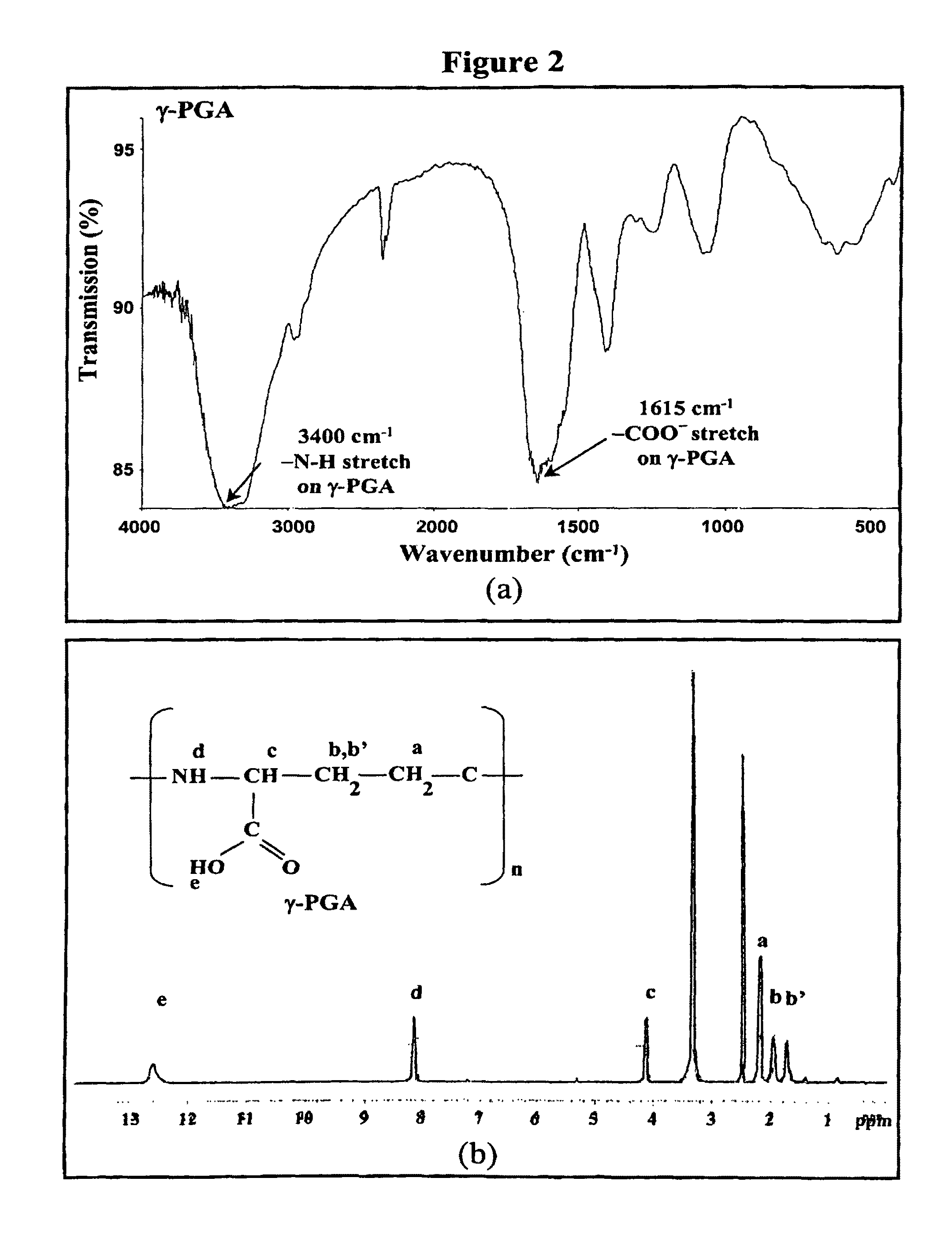
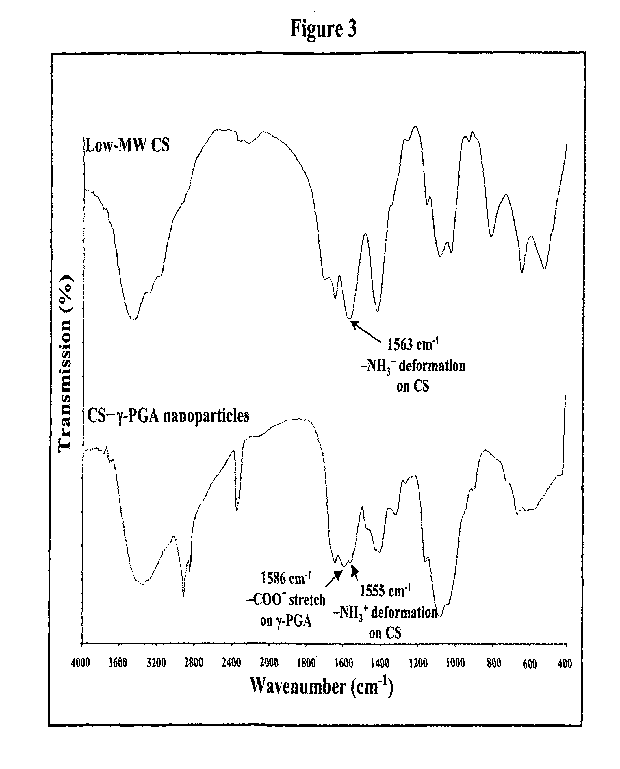
[0072]Nanoparticles were obtained upon addition of γ-PGA aqueous solution (pH 7.4, 2 ml), using a pipette (0.5-5 ml, PLASTIBRAND®, BrandTech Scientific Inc., Germany), into a low-MW CS aqueous solution (pH 6.0, 10 ml) at varying concentrations (0.01%, 0.05%, 0.10%, 0.15%, or 0.20% by w / v) under magnetic stirring at room temperature. Nanoparticles were collected by ultracentrifugation at 38,000 rpm for 1 hour. Supernatants were discarded and nanoparticles were resuspended in deionized water for further studies. The nanoparticles thus obtained via the simple and mild ionic-gelation method described herein show typical characteristics in a spheroidal configuration with a particle size of between about 50 to 400 nm, a positive surface charge and a narrow polydispersity index. FT-IR was used to analyze peak variations of amino groups of low-MW CS and carboxylic acid salts of γ-PGA in the CS-γ-PGA nanoparticles.

[0073]As stated, nanopar...


PUM

PropertyMeasurementUnit
diameteraaaaaaaaaa
MWaaaaaaaaaa
MWaaaaaaaaaa

Abstract

The invention discloses the bioactive nanoparticles composed of chitosan, poly-glutamic acid, and at least one bioactive agent characterized with a positive surface charge and enhanced permeability in oral drug delivery.

Description

CROSS-REFERENCE TO RELATED APPLICATIONS[0001]This application is a continuation-in-part application of U.S. patent application Ser. No. 12 / 286,504, filed Sep. 30, 2008, now U.S. Pat. No. 7,604,795, which is a continuation-in-part application of U.S. patent application Ser. No. 12 / 151,230, filed May 5, 2008, now U.S. Pat. No. 7,541,046, which is a continuation-in-part application of U.S. patent application Ser. No. 11 / 398,145, filed Apr. 5, 2006, now U.S. Pat. No. 7,381,716, which is a continuation-in-part application of U.S. patent application Ser. No. 11 / 284,734, filed Nov. 21, 2005, now U.S. Pat. No. 7,282,194, which is a continuation-in-part application of U.S. patent application Ser. No. 11 / 029,082, filed Jan. 4, 2005, now U.S. Pat. No. 7,265,090, the entire contents of all are incorporated herein by reference. This application also claims priority benefits of provisional patent application Ser. No. 61 / 204,025, filed Dec. 31, 2008, the entire contents of which are incorporated h...

Claims


Application Information

Patent Timeline
Patent Type & Authority Patents(United States)
IPC IPC(8): A61K51/00A61K9/50
CPCA61K47/36A61K9/2077A61K9/2081A61K9/2806A61K9/4891A61K9/5146A61K9/5161A61K9/5192A61K31/13A61K31/445A61K31/55A61K47/26A61K47/34A61K9/19A61K9/0043
Inventor SUNG, HSING-WENTU, HOSHENG
Owner NANOMEGA MEDICAL CORP
Why Eureka
Try Eureka