
Human binding molecules having killing activity against staphylococci and uses thereof

a technology of staphylococcus and binding molecules, which is applied in the field of medicine, can solve the problems of major medical and consequent economic problems, pharmaceutical companies appear to have lost interest in the antibiotic market, and so as to prevent, cure or stop or inhibit the spread of staphylococcus, and the effect of reducing the cost of new drug developmen

Active Publication Date: 2012-07-03
JANSSEN VACCINES & PREVENTION BV
View PDF144 Cites 33 Cited by
  • Summary
  • Abstract
  • Description
  • Claims
  • Application Information

AI Technical Summary

Benefits of technology

[0084]Alternatively, phage display libraries may be constructed from immunoglobulin variable regions that have been partially assembled in vitro to introduce additional antibody diversity in the library (semi-synthetic libraries). For example, in vitro assembled variable regions contain stretches of synthetically produced, randomized or partially randomized DNA in those regions of the molecules that are important for antibody specificity, e.g. CDR regions. Phage antibodies specific for bacteria such as staphylococci can be selected from the library by exposing the bacteria or material thereof to a phage library to allow binding of phages expressing antibody fragments specific for the bacteria or material thereof. Non-bound phages are removed by washing and bound phages eluted for infection of E. coli bacteria and subsequent propagation. Multiple rounds of selection and propagation are usually required to sufficiently enrich for phages binding specifically to the bacteria or material thereof. If desired, before exposing the phage library to the bacteria or material thereof the phage library can first be subtracted by exposing the phage library to non-target material such as bacteria of a different family, species and / or strain or bacteria in a different growth phase or material of these bacteria. These subtractor bacteria or material thereof can be bound to a solid phase or can be in suspension. Phages may also be selected for binding to complex antigens such as complex mixtures of bacterial proteins or (poly)peptides optionally supplemented with bacterial polysaccharides or other bacterial material. Host cells expressing one or more proteins or (poly)peptides of bacteria such as staphylococci may also be used for selection purposes. A phage display method using these host cells can be extended and improved by subtracting non-relevant binders during screening by addition of an excess of host cells comprising no target molecules or non-target molecules that are similar, but not identical, to the target, and thereby strongly enhance the chance of finding relevant binding molecules. Of course, the subtraction may be performed before, during or after the screening with bacterial organisms or material thereof. The process is referred to as the Mabstract® process (Mabstract® is a registered trademark of Crucell Holland B.V., see also U.S. Pat. No. 6,265,150 which is incorporated herein by reference).
[0094]Alternatively, the binding molecules, immunoconjugates, nucleic acid molecules or compositions of the present invention can be in solution and the appropriate pharmaceutically acceptable excipient can be added and / or mixed before or at the time of delivery to provide a unit dosage injectable form. Preferably, the pharmaceutically acceptable excipient used in the present invention is suitable to high drug concentration, can maintain proper fluidity and, if necessary, can delay absorption.
[0099]In a further aspect, the binding molecules such as human monoclonal antibodies (functional fragments and variants thereof), immunoconjugates, compositions, or pharmaceutical compositions of the invention can be used as a medicament. So, a method of treatment and / or prevention of a bacterial (Gram-positive and / or Gram-negative), e.g. a staphylococcal, infection using the binding molecules, immunoconjugates, compositions, or pharmaceutical compositions of the invention is another part of the present invention. The above-mentioned molecules can inter alia be used in the diagnosis, prophylaxis, treatment, or combination thereof, of a bacterial infection. They are suitable for treatment of yet untreated patients suffering from a bacterial infection and patients who have been or are treated for a bacterial infection. They may be used for patients such as hospitalized infants, premature infants, burn victims, elderly patients, immunocompromised patients, immunosuppressed patients, patient undergoing an invasive procedure, and health care workers. Each administration may protect against further infection by the bacterial organism for up to three or four weeks and / or will retard the onset or progress of the symptoms associated with the infection. The binding molecules of the invention may also increase the effectiveness of existing antibiotic treatment by increasing the sensitivity of the bacterium to the antibiotic, may stimulate the immune system to attack the bacterium in ways other than through opsonization. This activation may result in long lasting protection to the infection bacterium. Furthermore, the binding molecules of the invention may directly inhibit the growth of the bacterium or inhibit virulence factors required for its survival during the infection.
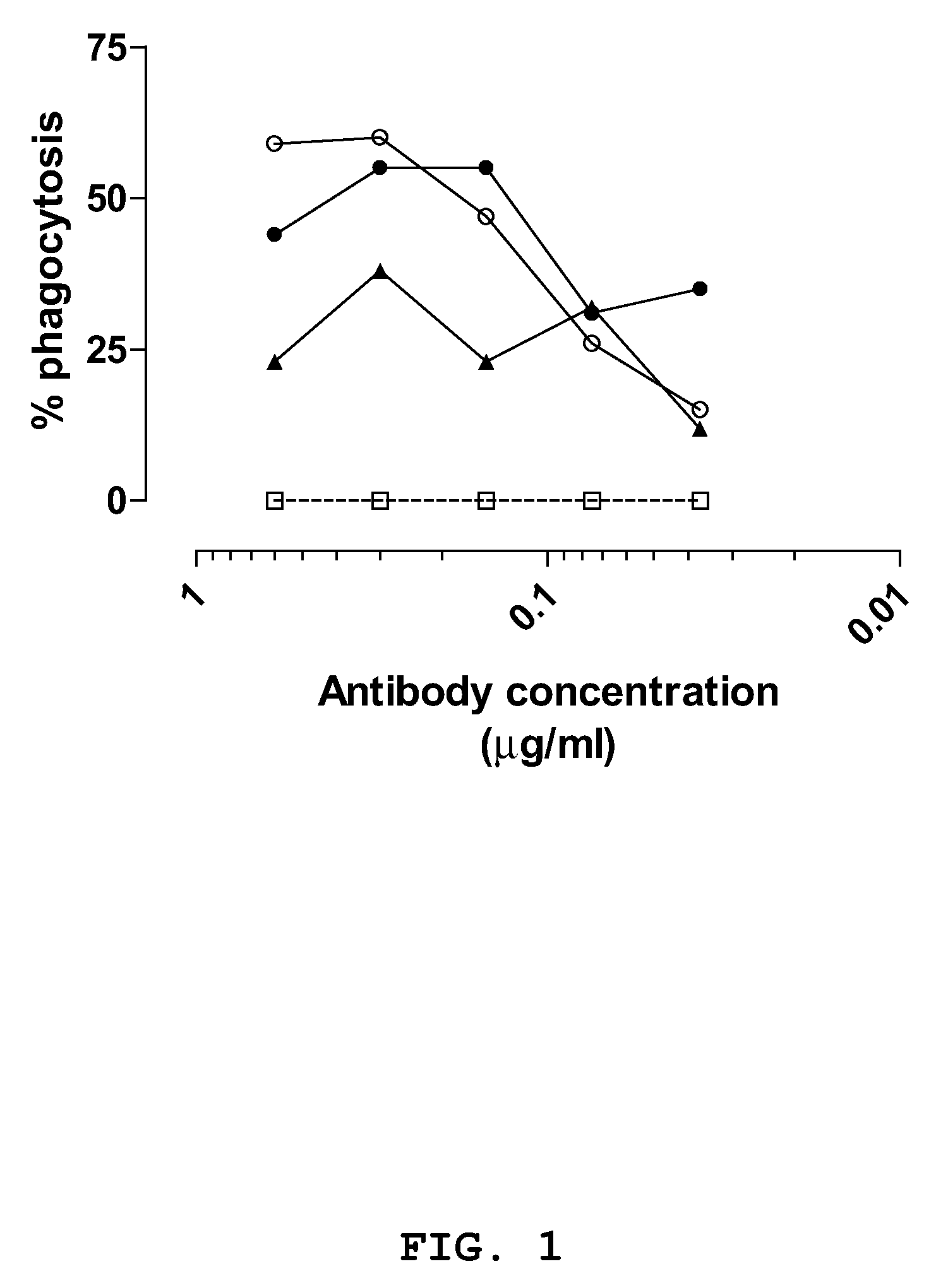
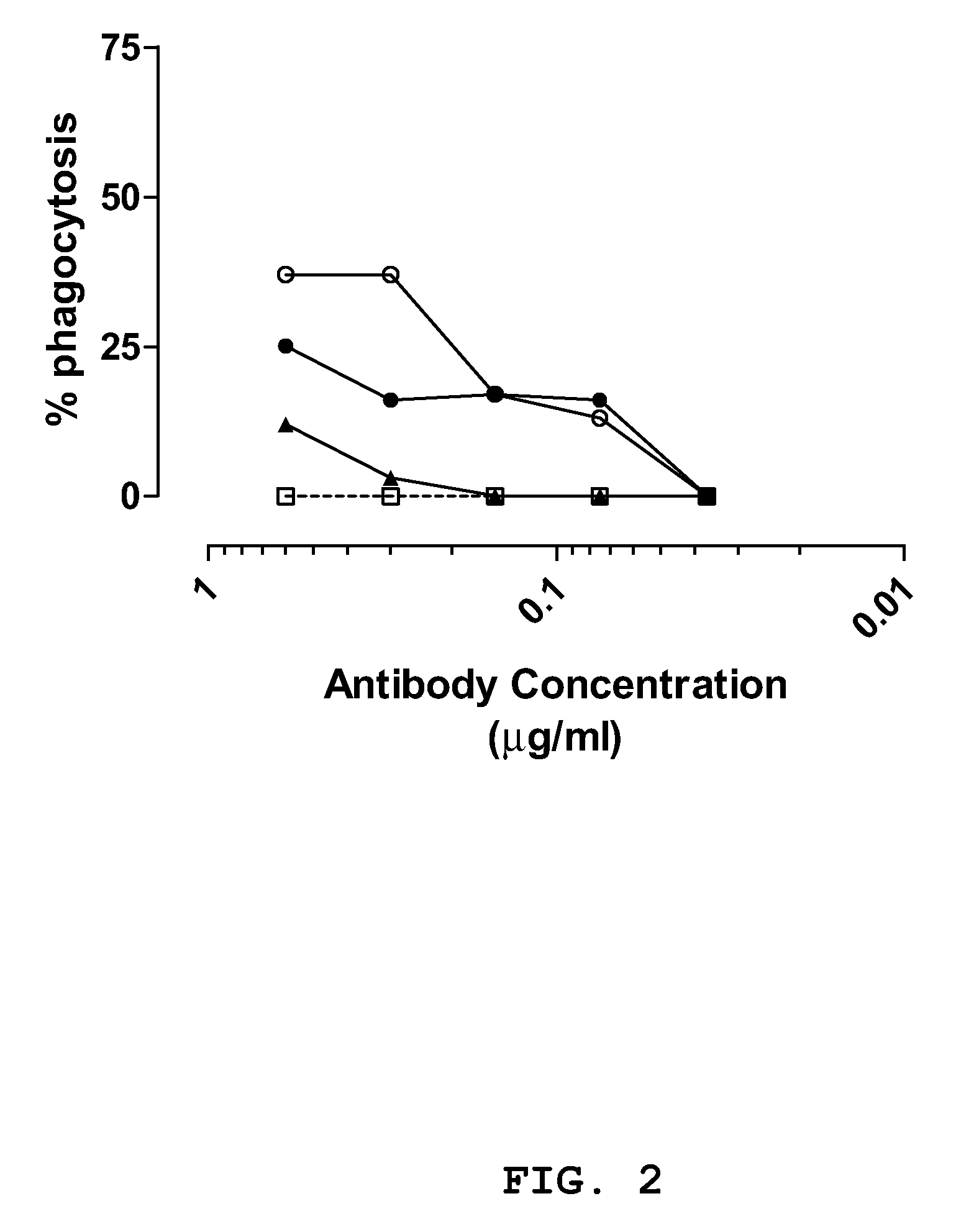
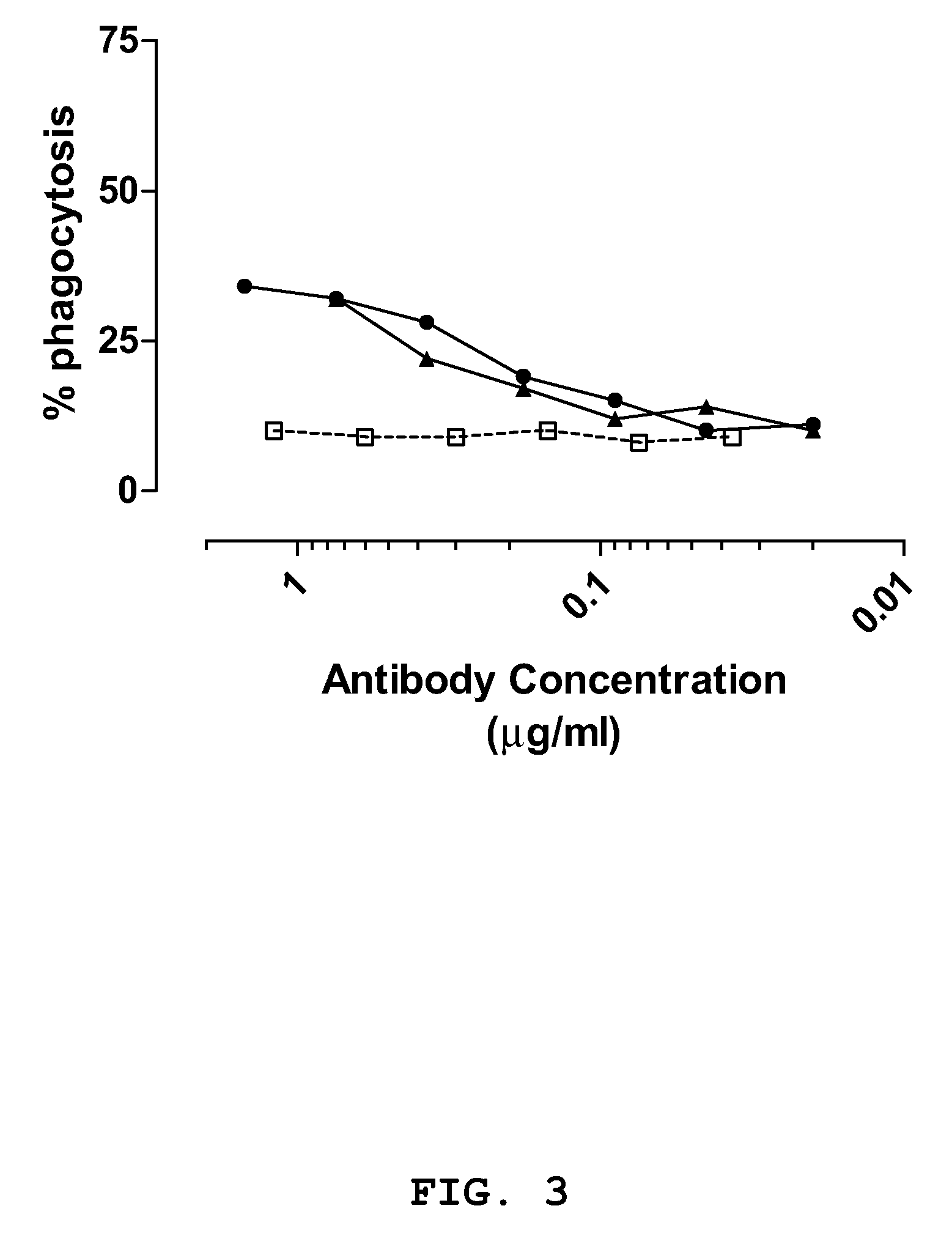
[0130]The anti-staphylococcal IgGs and a control IgG (CR4374) were serially diluted in opsonophagocytosis buffer in a total volume of 20 μl to obtain dilutions having an IgG concentration of 2.50 μg / ml, 1.20 μg / ml, 0.60 μg / ml, 0.30 μg / ml, 0.15 μg / ml, 0.075 μg / ml, 0.0375 μg / ml and 0.019 μg / ml. Opsonic activity of dilutions was measured in the OPA assay in a round bottom plate that was blocked with 1% BSA in PBS. As a control, the assay was performed with no IgG. A 15 μl aliquot of a bacterial suspension containing 5.4×10⁶ cells was added to each well of the plate. When a bacterial suspension from S. aureus strain Cowan or S. epidermidis was used, the IgG / bacterium suspension was first incubated for 30 minutes at 37° C. while the plate was horizontally shaking (1300 rpm) in a Heidolph titramax 1000. Next, 15 μl of the differentiated HL-60 cells (total: 75×103 cells) were added to each well of the plate and the plate was incubated while shaking at 37° C. for 30-45 minutes. The final volume in the well was 50 μl. The reaction was stopped by adding 50 μl of wash buffer containing 4% v / v formaldehyde. The content in each well was resuspended and transferred to polystyrene disposable tubes for flow cytometric analysis. The samples were stored in the dark at 4° C. until analyzation. The tubes were vortexed for 3 seconds before sampling in the flow cytometer. To control the differentiation of the HL-60 cells the expression of the complement receptor CD11b was measured. Fc-receptors of differentiated and non-differentiated cells were first blocked with rabbit IgG for 15 minutes on ice and the cells were subsequently labelled with CD11bAPC (BD) for 15 minutes on ice. Cells were considered properly differentiated when the mean fluorescent intensity (MFI) analyzed was at least between 10- to 100-fold higher compared to that of non-differentiated cells. Samples were assayed with a FACSCalibur immunocytometry system (Becton Dickinson and Co., Paramus, N.J.) and were analyzed with CELLQuest software (version 1.2 for Apple system 7.1; Becton Dickinson). 7,000 gated HL-60 granulocytes were analyzed per tube. FAM-SE was excited at a wavelength of 488 nm and the FAM-SE fluorescence signal of gated viable HL-60 cells was measured for each antibody dilution. IgGs were defined as positive in the phagocytic assay when concentration dependent phagocytosis could be observed greater or equal to two times that of the control IgG. IgGs CR2430, CR5132 and CR5133 demonstrated opsonic activity against S. aureus strain Cowan in both the log (see FIG. 1) and stationary growth phase (see FIG. 2). The three IgGs where more effective in enhancing phagocytic activity during the log phase of growth. IgGs CR5132 and CR5133 enhanced phagocytosis of S. aureus strain SA125 compared to the negative control antibody (see FIG. 3) and antibody CR5133 significantly enhanced phagocytic activity of the differentiated HL60 cells against S. epidermidis strain SE131, when compared to the negative control antibody (see FIG. 4).Example 8Breadth of Staphylococci Specific IgG1 Binding Activity

Problems solved by technology

However, over the past 30 years, coagulase-negative staphylococcal infections have emerged as one of the major complications of medical progress.
Of the estimated 2 million hospital infections in the US in 2004, 70% was resistant to at least one antibiotic, thereby causing major medical and consequently economic problems.
Despite the urgent need for the development of new antibiotics, the major pharmaceutical companies appear to have lost interest in the antibiotic market.
This trend has been attributed to several factors: the cost of new drug development and the relatively small return on investment that infectious disease treatments yield compared to drugs against hypertension, arthritis and lifestyle drugs e.g. for impotence.
Another contributing factor is the increasing difficulty in finding new targets, further driving up development costs.
As a consequence thereof, vaccines are very complex, take long and are expensive to develop.
The immune status of the populations at risk for nosocomial infections further complicates vaccine design.
These patients are inherently unwell and may even be immunocompromised (due to the effect of immunosuppressive drugs) resulting in delayed or insufficient immunity against the infecting pathogens.
Furthermore, except in the case of certain elective procedures, it may not be possible to identify and vaccinate the at risk patients in time to give them sufficient immune protection from infection.
However, blood-derived products have potential health risks inherently associated with these products.
In addition, the immunoglobulins can display batch-to-batch variation and may be of limited availability in case of sudden mass exposures.
However, murine antibodies are limited for their use in vivo due to problems associated with administration of murine antibodies to humans, such as short serum half life, an inability to trigger certain human effector functions and elicitation of an unwanted dramatic immune response against the murine antibody in a human (HAMA).
A disadvantage of these chimeric antibodies is however that they still retain some murine sequences and therefore still elicit an unwanted immune reaction, especially when administered for prolonged periods.

Method used


Image

  • Human binding molecules having killing activity against staphylococci and uses thereof
  • Human binding molecules having killing activity against staphylococci and uses thereof
  • Human binding molecules having killing activity against staphylococci and uses thereof

Examples


example 1

Construction of scFv Phage Display Libraries Using RNA Extracted from Donors Screened for Opsonic Activity

[0113]Samples of blood were taken from donors reporting a recent gram-positive bacterial infection as well as healthy adults between 25-50 years of age. Peripheral blood leukocytes were isolated by centrifugation and the blood serum was saved and frozen at −80° C. Donor serum was screened for opsonic activity using a FACS-based phagocytosis assay (Cantinieaux et al., 1989) and compared to a pool of normal healthy donor serum. Sera from donors having a higher phagocytic activity compared to normal serum were chosen to use for the generation of phage display libraries. Total RNA was prepared from the peripheral blood leukocytes of these donors using organic phase separation and subsequent ethanol precipitation. The obtained RNA was dissolved in RNAse-free water and the concentration was determined by OD 260 nm measurement. Thereafter, the RNA was diluted to a concentration of 100 ...

example 2

Construction of scFv Phage Display Libraries Using RNA Extracted from Memory B Cells

[0116]Peripheral blood was collected from normal healthy donors, convalescent donors or vaccinated donors by venapunction using EDTA anti-coagulation sample tubes. A blood sample (45 ml) was diluted twice with PBS and 30 ml aliquots were underlayed with 10 ml Ficoll-Hypaque (Pharmacia) and centrifuged at 900×g for 20 minutes at room temperature without breaks. The supernatant was removed carefully to just above the white layer containing the lymphocytic and thrombocytic fraction. Next, this layer was carefully removed (˜10 ml), transferred to a fresh 50 ml tube and washed three times with 40 ml PBS and spun at 400×g for 10 minutes at room temperature to remove thrombocytes. The obtained pellet containing lymphocytes was resuspended in RPMI medium containing 2% FBS and the cell number was determined by cell counting. Approximately 1×10⁸ lymphocytes were stained for fluorescent cell sorting using CD24,...

example 3

Selection of Phages Carrying Single Chain Fv Fragments Specifically Binding to Staphylococci

[0117]Antibody fragments were selected using antibody phage display libraries, general phage display technology and MAbstract® technology, essentially as described in U.S. Pat. No. 6,265,150 and in WO 98 / 15833 (both of which are incorporated by reference herein). The antibody phage libraries used were screened donor libraries prepared as described in Example 1, IgM memory libraries prepared as described in Example 2 and a semi-synthetic scFv phage library (JK1994) which has been described in de Kruif et al., 1995b. The methods and helper phages as described in WO 02 / 103012 (incorporated by reference herein) were used in the present invention. For identifying phage antibodies recognizing staphylococci, phage selection experiments were performed using live bacteria in suspension. The clinical isolates used for selection and screening are described in Table 8. The isolates are different based on...


PUM


Abstract

The present invention provides human binding molecules specifically binding to staphylococci and having killing activity against staphylococci, nucleic acid molecules encoding the human binding molecules, compositions comprising the human binding molecules and methods of identifying or producing the human binding molecules. The human binding molecules can be used in the diagnosis, prophylaxis and / or treatment of a condition resulting from Staphylococcus.

Description

CROSS-REFERENCE TO RELATED APPLICATION[0001]This application is the national phase entry of PCT International Patent Application No. PCT / EP2007 / 055527, filed on Jun. 5, 2007, designating the United States of America, and published, in English, as PCT International Publication No. WO 2007 / 141274 A2 on Dec. 13, 2007, which itself claims the benefit of U.S. Provisional Patent Application Ser. No. 60 / 811,477, filed Jun. 6, 2006, EP 06124231.9, filed Nov. 16, 2006, and EP 07103584.4 filed on Mar. 6, 2007.FIELD OF THE INVENTION[0002]The invention relates to medicine. In particular the invention relates to the diagnosis, prophylaxis and / or treatment of infection by staphylococci.BACKGROUND OF THE INVENTION[0003]Staphylococcus is a genus of gram-positive bacteria and a member of the micrococcaceae family. Staphylococci are spherical bacteria that are found primarily on the skin and in the mucous membranes of humans and other warm-blooded animals, and aggregate into small, grape-like clumps....

Claims


Application Information

Patent Timeline
Patent Type & Authority Patents(United States)
IPC IPC(8): A61K39/40
CPCC07K16/1271A61K2039/505C07K2317/21C07K2317/77C07K2317/565C07K2317/622C07K2317/56A61P31/00A61P31/04A61P43/00C07K16/12A61K39/40C12N15/11
Inventor THROSBY, MARKGEUIJEN, CECILIA A. W.DE KRUIF, CORNELIS ADRIAAN
Owner JANSSEN VACCINES & PREVENTION BV
Learn More