Method for detecting bacteria
a colorimetric assay and bacteria technology, applied in the field of colorimetric assay and method, can solve the problems of no urine colorimetric assay, laborious and time-consuming, and significant levels of bacterial growth, and achieve the effects of ensuring the safety of platelets, rapid, sensitive and specific detection, and ensuring the safety of bacterial populations
- Summary
- Abstract
- Description
- Claims
- Application Information
AI Technical Summary
Benefits of technology
Problems solved by technology
Method used
Image
Examples
example 1
Bacteria Spiked Platelets
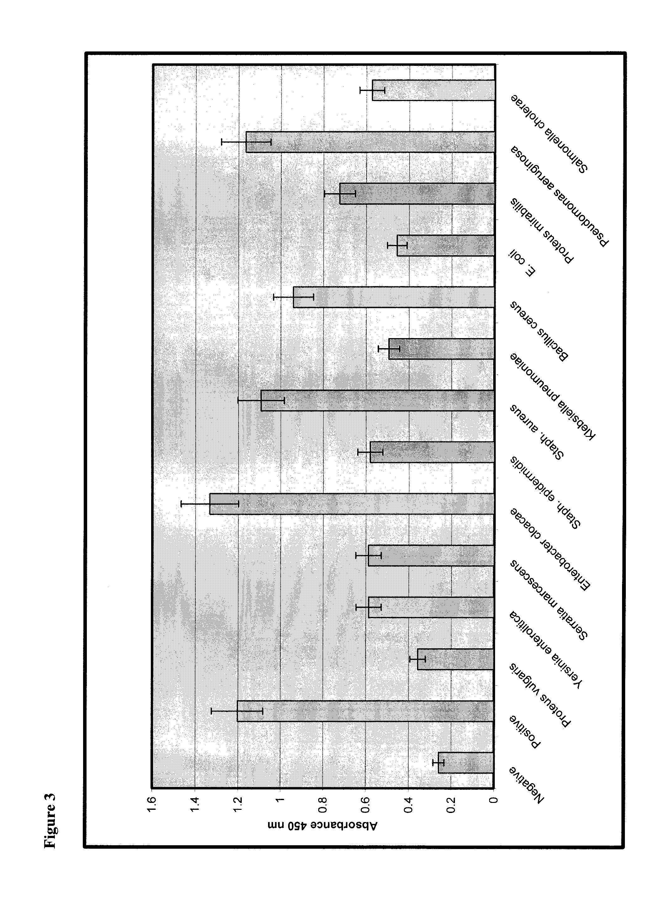
[0110]In order to assess the sensitivity of the assay, spiking studies were performed with the following bacteria species: Proteus vulgaris, Yersinia enterocolitica, Serratia marcescens (S. marcescens), Enterobacter cloacae, Staphylococcus epidermidis (S. epidermidis), Staphylococcus aureus (S. aureus), Klebsiella pneumoniae, Bacillus cereus, Escherichia coli (E. coli), Proteus mirabilis, Pseudomonas aeruginosa (P. aeruginosa), and Salmonella cholerae. All of these species represent common skin flora. Bacteria species were obtained from the American Type Culture Collection.
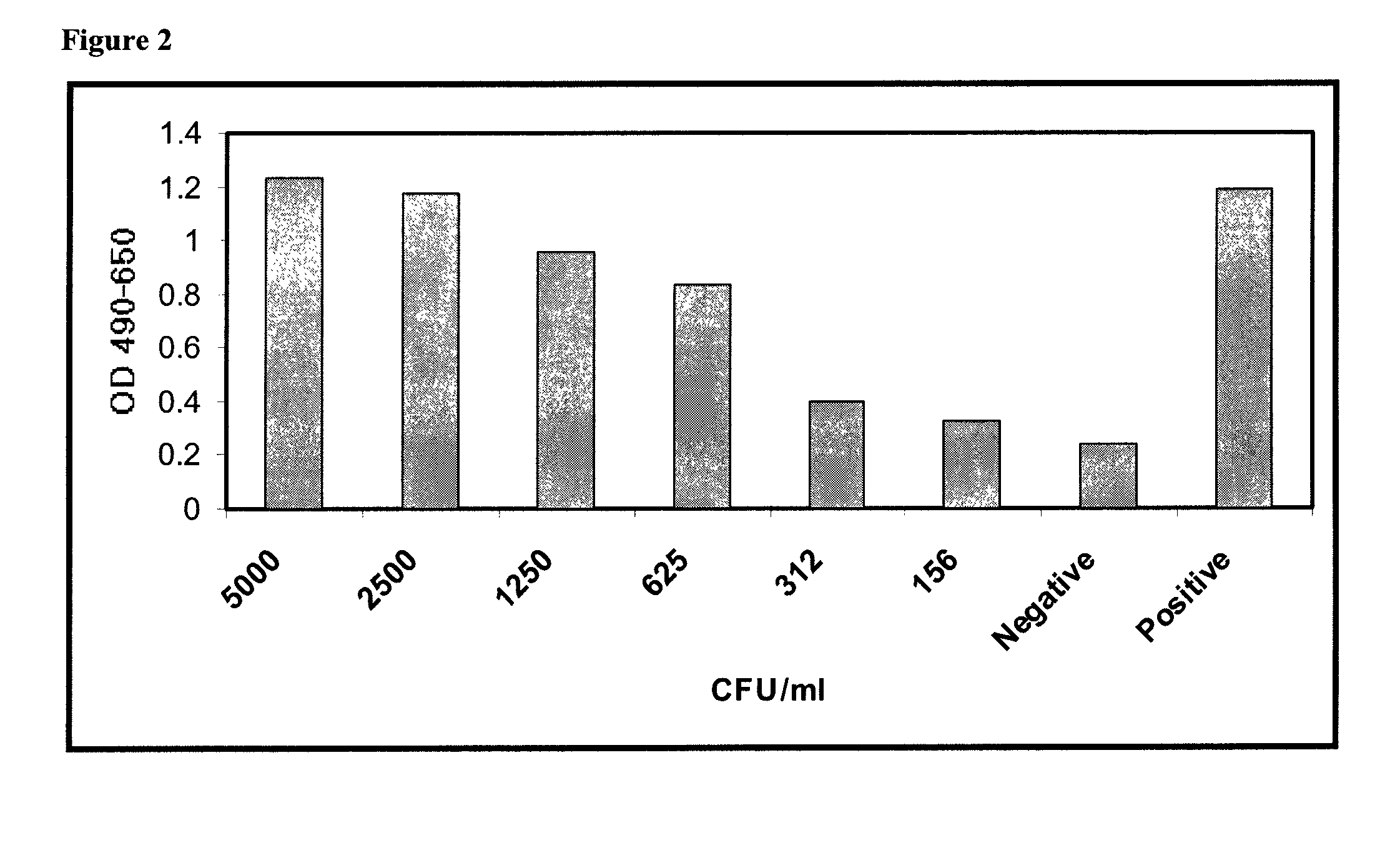
[0111]Bacteria were cultured and quantified by reconstituting the bacteria according to the vendor's instructions and grown in Trypticase Soy Broth (Becton Dickinson) overnight at 37° C. Bacteria were washed twice by centrifugation in sterile phosphate buffered saline (PBS) and resuspended in 5 ml sterile PBS. Serial 10-fold dilutions were prepared in PBS by adding 100 μl bacterial suspens...
example 2
Time Course Study of Bacterial Growth in Platelets
[0117]In a different approach to assessment of assay sensitivity, a time course study of bacterial growth in platelets was performed using Assay Method 2. In this series of experiments, assay results vs. CFU / ml were determined at increasing times following inoculation of a platelet bag with bacteria. Platelets from two units were removed from platelet bags and placed in sterile 50 ml conical tubes for ease of sampling over time. At time zero, one unit was spiked with 10 CFU / ml of S. aureus (indicated as SA in FIG. 8), and the other was spiked at 10 CFU / ml of P. aeruginosa (indicated as PA in FIG. 8). Immediately after spiking, one ml samples were taken from each. Following incubation for 20 hours and 48 hours, one ml samples were taken from each. Immediately upon harvest, samples were placed at 4° C. to inhibit further bacterial growth until the time of assay. All samples were processed in parallel as follows: one ml of each was cent...
example 3
Specificity of the Assay for Peptidoglycan
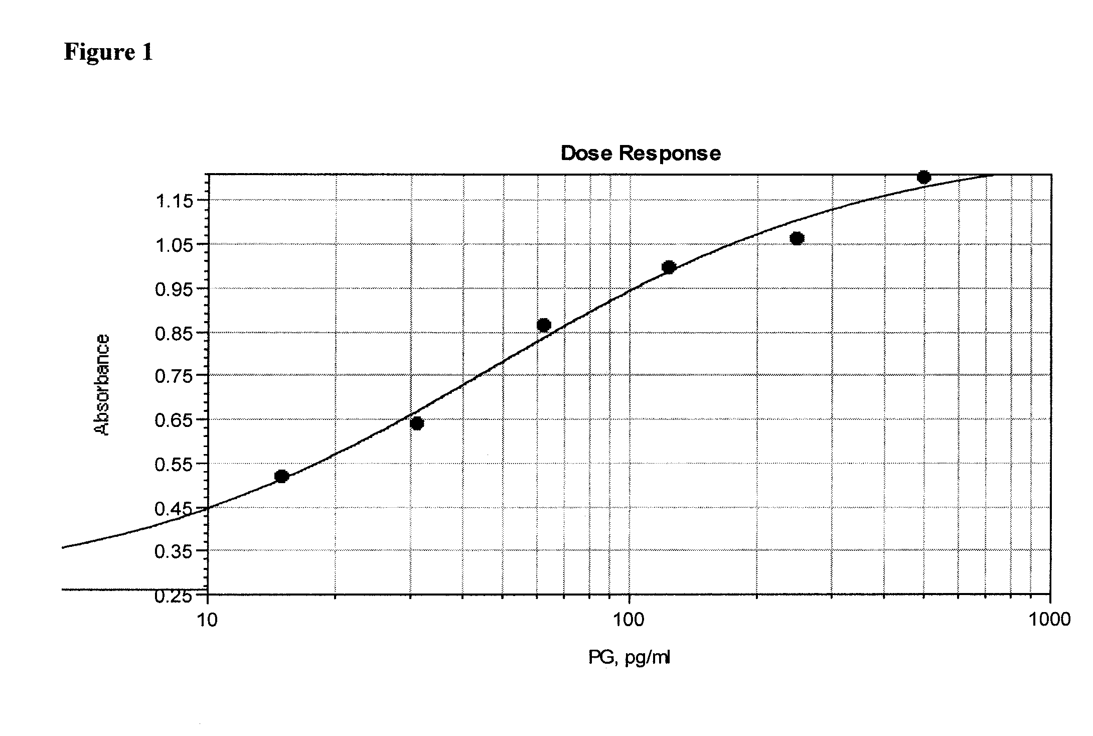
[0119]In order to demonstrate the specificity of the assay for peptidoglycan, a dose response curve was constructed by serially diluting purified peptidoglycan at concentrations from 10 ng / ml to 150 pg / ml into extracted and neutralized platelets as described in Assay Method 1. The absorbance at 490 nm, corrected for background read at 650 nm, is proportional to the peptidoglycan concentration (FIG. 10). A lower limit of 156 pg / ml peptidoglycan was visually detectable.
PUM
| Property | Measurement | Unit |
|---|---|---|
| concentrations | aaaaa | aaaaa |
| wavelengths | aaaaa | aaaaa |
| time | aaaaa | aaaaa |
Abstract
Description
Claims
Application Information
 - R&D
- Intellectual Property
- Life Sciences
- Materials
- Tech Scout
- Unparalleled Data Quality
- Higher Quality Content
- 60% Fewer Hallucinations
Browse by: Latest US Patents, China's latest patents, Technical Efficacy Thesaurus, Application Domain, Technology Topic, Popular Technical Reports.
© 2025 PatSnap. All rights reserved.Legal|Privacy policy|Modern Slavery Act Transparency Statement|Sitemap|About US| Contact US: help@patsnap.com


