
Molecular probes for multimodality imaging and tracking of stem cells

a multi-modality, stem cell technology, applied in the field of imaging probes, can solve the problems of label efflux from cells, cell death, cell death, etc., and achieve the effect of accurate determination of stem cell biodistribution

Active Publication Date: 2016-11-15
UNIV OF MASSACHUSETTS
View PDF1 Cites 0 Cited by
  • Summary
  • Abstract
  • Description
  • Claims
  • Application Information

AI Technical Summary

Benefits of technology

The invention describes a new type of molecular probe that can be used to image stem cells in vivo using both fluorescence and radionuclide imaging techniques. These probes have a unique structure that allows them to attach securely to the cell membrane, preventing the labels from leaching out of the cells. This results in a more accurate determination of stem cell biodistribution and improved sensitivity. The probes are also minimally toxic, allowing for a greater number of radiolabels per cell.

Problems solved by technology

One of the concerns with stem cell therapies is the risk that transplanted stem cells could form tumors and become cancerous if cell division continues uncontrollably.
It has two inherent limitations: labels may be diluted upon cell division, making the cells eventually invisible; and labels may efflux from cells or may degrade over time.
Major disadvantages of the reporter gene approach include: (i) stable transfection (lentiviral or retroviral) involves extensive molecular manipulation of the cells under study and runs the risk of insertional mutagenesis; (ii) immune reactions may be induced; (iii) uncertainties regarding the robustness of the signal remain, as the detected signal could reflect magnitude of transgene expression instead of cell survival; (iv) gene modification adds additional cost; and (v) regulatory roadblocks.
Another significant clinical problem common to all MRI methods is that certain implantable devices such as pacemakers and defibrillators, are currently contraindications to MRI.
Current radioisotope and optical probes for stem cell tracking have limitations.
However, this approach suffers from many of the same limitations previously mentioned.
For stem cell tracking, the short radiological half-life of 99mTc (6.02 h) is not particularly useful for longer term imaging studies using the direct radiolabeling approach.
One problem reported with cells labeled with 111In-oxine or 111In-tropolone is the potential for accumulation of the isotope in the nucleus, resulting in radiotoxicity and limiting the amount of label possible per cell.
This is due to the short range of the emitted low-energy B-particles, causing severe chromosomal aberration.
Detection at the single cell level remains a formidable challenge for radionuclide probes as the ability to concentrate radioactive agents in stem cells has yet to be achieved.
Another important limitation that has been reported with 111In-oxine and 111In-tropolone is the leaking of the label from the stem cell resulting in false positive signals and also high uptake in liver and kidneys.
The use of an iodine isotope presents practical issues.
First, exchange labeling is not efficient in producing a high specific radioactivity agent.
Second, it is challenging to conform to a simple kit format that can be easily radiolabeled on site.

Method used


Image

  • Molecular probes for multimodality imaging and tracking of stem cells
  • Molecular probes for multimodality imaging and tracking of stem cells
  • Molecular probes for multimodality imaging and tracking of stem cells

Examples


example 1

Preparation of Exemplary Compounds (as Shown in Scheme 1)

[0136]Preparation of Compound (1):

[0137]Compound (1) was prepared according to the procedure of Gale et al. (1977).

[0138]Preparation of Compound (2):

[0139]Compound (1) (20.6 g, 0.065 mol) and n-docosanyl-4-chlorobenzenesulfonate (32.4 g, 0.065 mol) were heated together at 130° C. with stirring for 2 h. The mixture was then cooled to RT and the crude solid recrystallized from ethyl acetate to yield pure (2) (36.4 g, 69%). 1H NMR (CDCl3, 200 MHz): δ7.50-7.90 (m, 8H), 7.20-7.30 (m, 3H), 4.95 (s, 2H), 4.65 (t, 2H), 2.95 (s, 3H), 1.7-1.9 (m), 1.55 (s, 6H), 1.10-1.40 (m), 0.85 (t, 3H).

[0140]Preparation of Compound (3):

[0141]Compound (2) (36 g, 0.044 mol) was dissolved in concentrated hydrochloric acid (660 mL) and the solution slowly heated to 115° C. over 2 h and then refluxed at this temperature for 22 h. The solution was then cooled to RT and placed in and ice bath and then taken to pH=9 with 30% ammonium hydroxide solution. The ...

example 2

Preparation of Example Compounds (as Shown in Scheme 2)

[0160]The amine functionalized dye (8) was reacted with glutaric anhydride to provide (12). Standard coupling of (12) with 2-(4-aminobenzyl) diethylenetriamine penta-t-butyl acetate (www.macrocyclics.com) using HATU provided (13) which was purified via column chromatography and isolated in high yield. Removal of the t-butyl esters from (13) was accomplished by treating with TFA in dichloromethane to furnish the free acid (14). (14) was used to prepare both natural and indium-111 labeled (15) using methods similar to those described above for labeling (10).

[0161]

[0162]Scheme 2 shows synthetic scheme for preparing non-radioactive and radioactive indium labeled compounds of the invention with a DTPA chelator. Reagents: (i) glutaric anhydride (ii) 2-(4-aminobenzyl) diethylenetriamine penta-t-butyl acetate, HATU (iii) TFA / CH2Cl2 (60:40) (iv) EtOH, natInCl3, 0.25M NaOAc or EtOH, 111InCl3, 0.25M NaOAc.

example 3

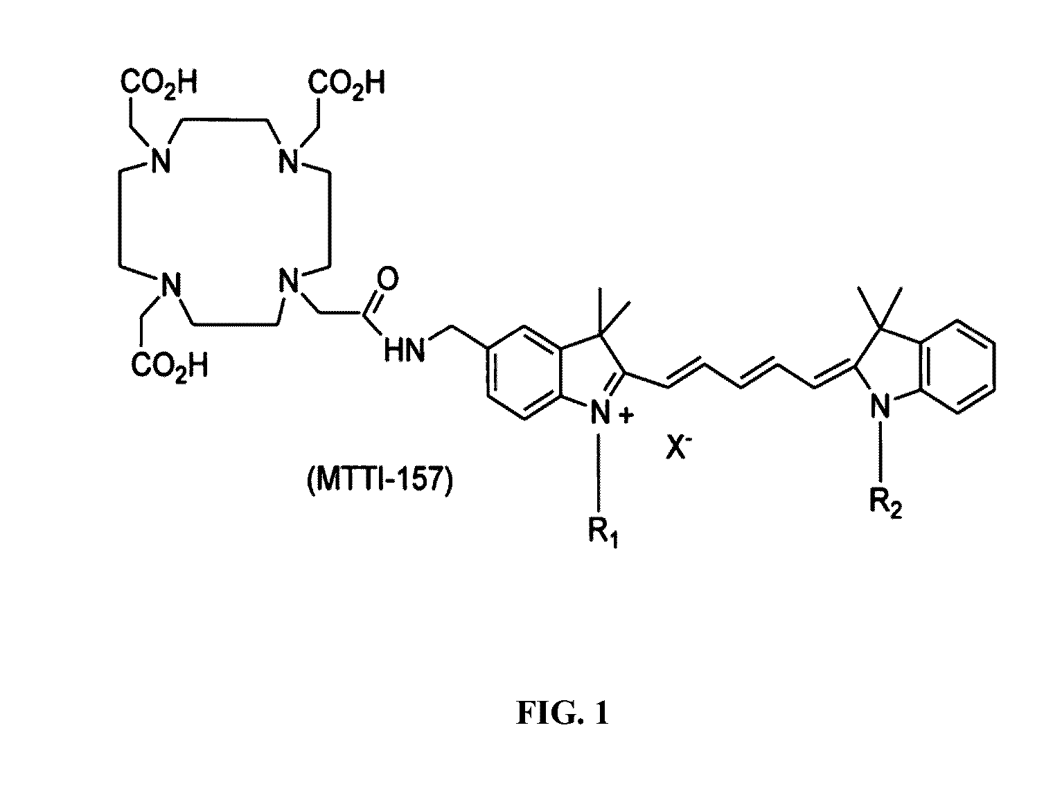
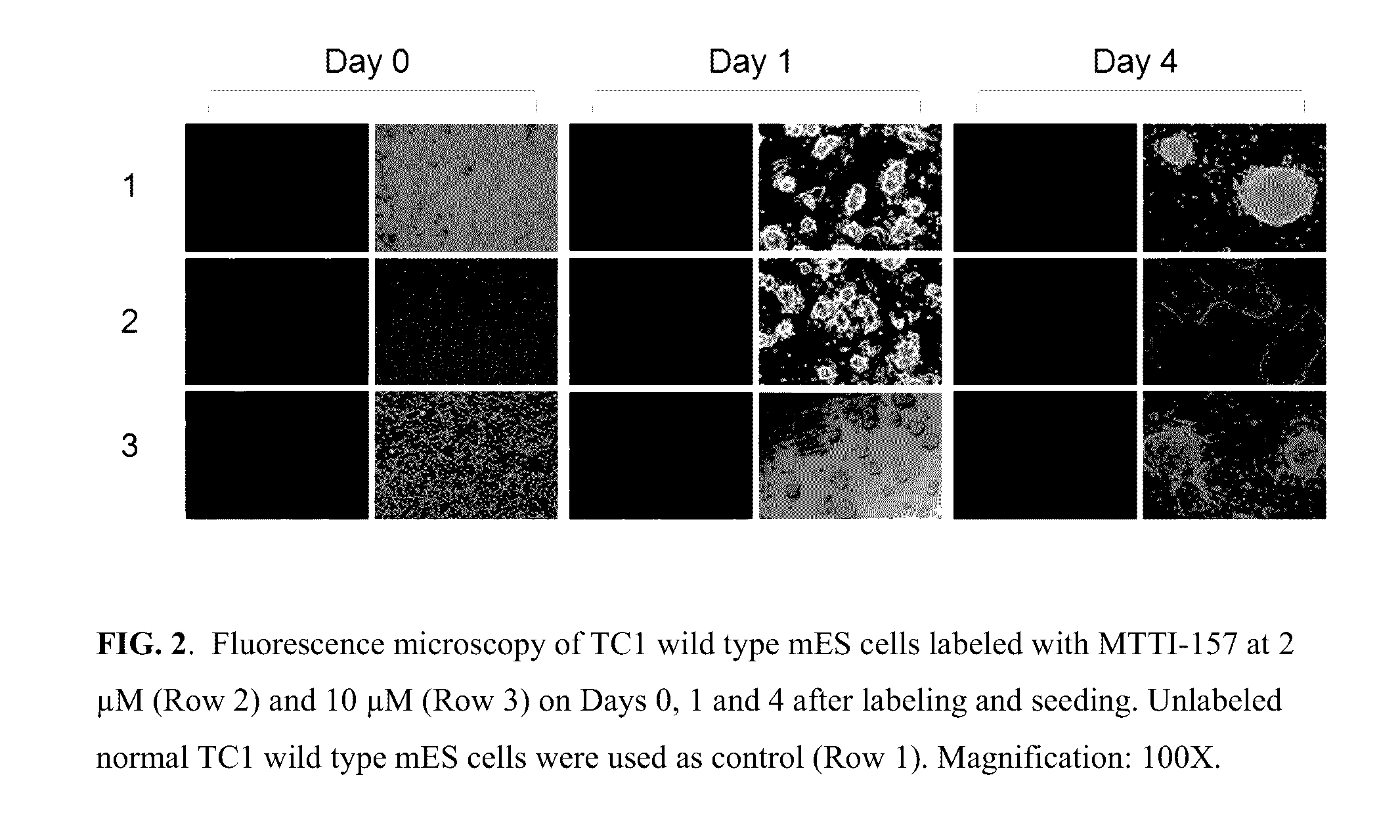
Stem Cells Labeling with MTTI-157 (No 111In)

[0163]TC1 wild type mouse embryonic stem (mES) cells were cultured in M15 medium (high glucose DMEM with 2 mM glutamine, 100 U / ml Pen / Strep, 0.1 mM beta mercaptoethanol, 15% FBS and 1000 U / ml murine leukemia inhibitory factor) on a freshly prepared gelatinized 100-mm tissue culture dish at 37° C. in a humidified atmosphere with 5% CO2. Medium was changed every day and cells passed every other day. For labeling with MTTI-157, the TC1 wild type mES cells were trypsinized, collected from the dishes, and washed twice with high glucose DMEM without serum. The washed cells were resuspended in Diluent C at the concentration of 2×10⁷ cells / ml. Immediately, prior to staining, a 2× working stock solution of MTTI-157 (4 μM, and 20 μM) was prepared in Diluent C. Then 1 ml of the cell suspension was added rapidly into 1 ml of the 2× working dye solution and immediately mixed by pipetting. The final staining solutions were 2 μM and 10 μM. After incubati...


PUM

PropertyMeasurementUnit
wavelengthsaaaaaaaaaa
temperatureaaaaaaaaaa
volumeaaaaaaaaaa

Abstract

The invention relates to novel multi-modality probes for imaging, tracking and analyzing stem cells and related biological samples, and methods of preparation and use thereof. The molecular probes of the invention are constructed, for example, by utilizing (a) the high selectivity of long hydrocarbon chains for binding to plasma membranes of cells, (b) a near-infrared (NIR) dye for optical imaging, and (c) a radionuclide for PET or SPECT imaging. The in vitro and in vivo data of the optical and radiolabeled probes demonstrated their utility for detecting the presence of stem cells with multiple imaging modalities.

Description

GOVERNMENT RIGHTS[0001]This invention was made with Government support under Grant No. 1R43GM093417-01 (SBIR) awarded by the National Institutes of Health. The United States Government has certain rights in the invention.PRIORITY CLAIMS AND RELATED PATENT APPLICATIONS[0002]This application claims the benefit of priority from and is the US national phase of International Application No. PCT / US13 / 42786, filed on May 27, 2013, which claims benefit of priority from U.S. Provisional Patent Application Ser. No. 61 / 654,794, filed Jun. 1, 2012, the entire content of each of which is incorporated herein by reference in its entirety for all purposes.TECHNICAL FIELDS OF THE INVENTION[0003]The invention generally relates to imaging probes. More particularly, the invention relates to novel multi-modality probes for imaging, tracking and analyzing stem cells and related biological samples, and methods of preparation and use thereof.BACKGROUND OF THE INVENTION[0004]Stem cell therapies have the pot...

Claims


Application Information

Patent Timeline
Patent Type & Authority Patents(United States)
IPC IPC(8): A61K51/04C07D403/14G01N33/50C07D209/20G01N33/58C07D209/14A61K49/00C07B59/00
CPCA61K51/0482A61K49/0032A61K51/0446A61K51/0478C07B59/002C07D209/14C07D209/20C07D403/14G01N33/5005G01N33/582A61K51/0485G01N33/52G01N33/56966
Inventor GRAY, BRIAN D.RUSCKOWSKI, MARYPAK, KOON Y.
Owner UNIV OF MASSACHUSETTS
Learn More