Biodegradable active medical tissue adhesive and its preparation method

A tissue adhesive and biodegradable technology, used in pharmaceutical formulations, surgical adhesives, applications, etc., can solve the problems of releasing toxic monomers, unfavorable to cells, and reducing the adsorption capacity of biocompatible calcium ions.

Inactive Publication Date: 2007-07-18
广州暨科拓源生物材料科技开发有限公司
View PDF0 Cites 12 Cited by
  • Summary
  • Abstract
  • Description
  • Claims
  • Application Information

AI Technical Summary

Problems solved by technology

[0008] (1) PMMA is used as the matrix phase, and some inherent shortcomings of PMMA cannot be avoided, such as difficulty in absorption, strong heat release during biocuring, release of toxic monomers, etc.
[0009] (2) The bio-inorganic filler is mainly artificially synthesized hydroxyapatite sintered at high temperature. The biocompatibility of HAP and t

Method used


Image

  • Biodegradable active medical tissue adhesive and its preparation method
  • Biodegradable active medical tissue adhesive and its preparation method

Examples


Embodiment 1

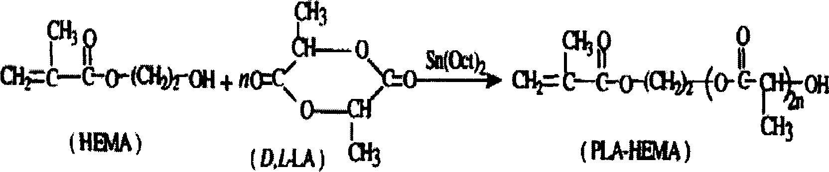
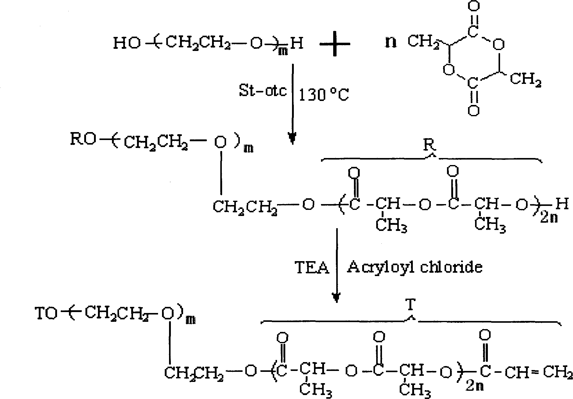
[0082] 0.22g macromonomer (MC-5), 0.34g degradable cross-linking agent were dissolved in 0.44g vinylpyrrolidone monomer, and 0.0075g benzoyl peroxide and 30ppm hydroquinone were added to obtain uniform and stable Clear Solution Component A.

[0083] When bonding skin lacerations, the operator first cleans up the wound thoroughly, without anesthesia and hemostasis, and uses the forefinger and thumb of the left hand to press lightly at a distance of 0.16 to 0.18 cm from the wound edge, which plays the role of oppression and hemostasis, and can Align the edges together. Drop 0.0050g of N,N-dimethyl-p-aniline into component A, mix well within 1min, and drop it on the wound so that it can completely cover the wound. After 2min to 3min, two fingers can slowly Loosen in a criss-cross pattern, and cover the wound with gauze after observing that the wound is no longer bleeding or splitting. After testing: the wound was completely absorbed and degraded after 8 weeks, and tissue sectio...

Embodiment 2

[0085] Dissolve 0.20g macromer (MC-3), 0.40g degradable cross-linking agent in 0.40g vinylpyrrolidone monomer, add 0.0075g benzoyl peroxide and 30ppm hydroquinone and mix to obtain uniform and stable Clear Solution Component A. In ophthalmic surgery, after the small and irregular corneal wound is sutured, add 0.005g N, N-dimethyl-p-aniline to component A, mix well within 1mim, drop into the wound that cannot be completely closed after suturing, 2min-3min Form a transparent film, which can effectively prevent the leakage of aqueous humor.

Embodiment 3

[0087] Dissolve 0.20g macromer (MC-5), 0.35g degradable cross-linking agent in 0.45g vinylpyrrolidone monomer, add 0.0075g benzoyl peroxide and 30ppm hydroquinone to obtain uniform and stable Clear solution.

[0088]New Zealand white rabbits were anesthetized and fixed in the supine position. According to the principle of aseptic technique, a midline incision was made on the upper abdomen to enter the abdominal cavity, and the upper part of the small intestine was cut off. Add 0.0050 g of N,N-dimethyl-p-aniline to the component one solution, and mix well within 1 min. Use it to glue the intestinal tube and the edge of the membrane. After 3 minutes, check that there is no leakage at the anastomosis, return the intestinal tube to the abdominal cavity, and close the abdomen. Absorption and degradation were basically complete after 12 weeks. Tissue sections showed that the material had good biocompatibility.


PUM


Abstract

A biodegradable and bioactive medical tissue adhesive with high bilcompatibility and adjustable mechanical strength and degradating speed for adhering soft tissue and hard tissue is prepared from the liquid phase (hydroxyethyl methylacrylate ring- opening polymerized cycloester type macro-molecular monomer), the alcohol type compound (biodegradable cross-linking agent, vinylpyrrolidone, N,N-dimethyl p- tolueneamine, or hydroquinone), and the solid phase (encapsulated natural powder, pore-forming agent, and benzoly peroxide) through mixing.

Description

technical field [0001] The invention relates to a medical adhesive, in particular to a biodegradable active medical tissue adhesive and a preparation method thereof. Background technique [0002] Human organs or tissues often lose their original functions due to injury or defect caused by trauma, inflammation, tumor, congenital malformation or surgery. Therefore, the research and development of medical adhesives suitable for various tissue repairs has become a global issue and is a very active field in biomaterials research. Existing medical adhesives can be divided into two categories, one is soft tissue adhesives, mainly α-hydroxyacrylate fast adhesives and fibrin adhesives; the other is hard tissue adhesives , mainly traditional methyl methacrylate-based bone cement and activated phosphate-based bone cement. [0003] Two kinds of soft tissue adhesives, a-hydroxyacrylate fast adhesive and fibrin adhesive, are widely used clinically, and have the advantages of fast adhesi...

Claims


Application Information

Patent Timeline
IPC IPC(8): A61L24/04A61K6/083
Inventor 周长忍杨媛罗丙红
Owner 广州暨科拓源生物材料科技开发有限公司
Why Eureka
Try Eureka