Vascular substrate without cell in vascular tissue and preparation method thereof

A decellularization and vascular technology, applied in the field of vascular matrix and its preparation, can solve the problems of graft failure, vascular intolerance to pressure conditions, unfavorable recipient cell infiltration and growth, etc., and achieve the effect of low cytotoxicity

Inactive Publication Date: 2009-07-08
XUANWU HOSPITAL OF CAPITAL UNIV OF MEDICAL SCI
View PDF0 Cites 27 Cited by
  • Summary
  • Abstract
  • Description
  • Claims
  • Application Information

AI Technical Summary

Problems solved by technology

[0004] Literature S. Kaushal, G.E. Amiel, K.J. Guleserian, O.M. Shapira, T. Perry, F.W. Sutherland, E. Rabkin, A.M. Moran, F.J. Schoen, A. Atala, S. Soker, J. Bischoff; and J.E. Mayer, Jr., Functional small-diameter neovessels created using endothelial progenitor cells expanded ex vivo. Nat Med7 (2001) 1035-40 provides such a method, but this method has an obvious defect: the cell components in the blood vessel wall cannot be removed cleanly, in practice In operation, after decellularization according to the method of this document, both HE staining and 4′, 6-diamidino-2-phenylindole (4′, 6-diamidino-2-phenylindole, DAPI) fluorescent staining can be used Seeing a large number of nuclei, cellular debris r

Method used


Image

  • Vascular substrate without cell in vascular tissue and preparation method thereof
  • Vascular substrate without cell in vascular tissue and preparation method thereof
  • Vascular substrate without cell in vascular tissue and preparation method thereof

Examples


Embodiment 1

[0046] Example 1 Preparation of decellularized vascular matrix

[0047] 1. Experimental materials:

[0048] 1) Blood vessel: the iliac artery of a freshly slaughtered pig taken from a nearby slaughterhouse, with a diameter of about 4-5 mm and a length of about 5 cm.

[0049] 2) Reagents: 1% Triton X-100 (Sigma); 0.1% Ammonium Hydroxide (Sigma); PBS (GIBCO); 0.25% Trypsin-0.02% EDTA (Sigma); 10% Fetal Bovine Serum (FBS, GIBCO) ; Low sugar-DMEM medium (LG-DMEM, GIBCO);

[0050] 3) Instruments: TS-100 decolorization shaker (Beijing Binda Yingchuang Technology Co., Ltd.); DK-8D constant temperature water tank (Shanghai Jinghong Experimental Equipment Co., Ltd.); LP-400 spectrophotometer (France); S-4800 Scanning electron microscope (HITACHI, Japan); AutoPore IV 9510 mercury porosimeter (Micromeritics Instruments Inc.USA) Shimadzu AG-5000A (Japan);

[0051] 2. Experimental method:

[0052] The blood vessels were taken from the adjacent slaughterhouse to take the iliac artery of...

Embodiment 2

[0059] Example 2 Detection of decellularized vascular matrix
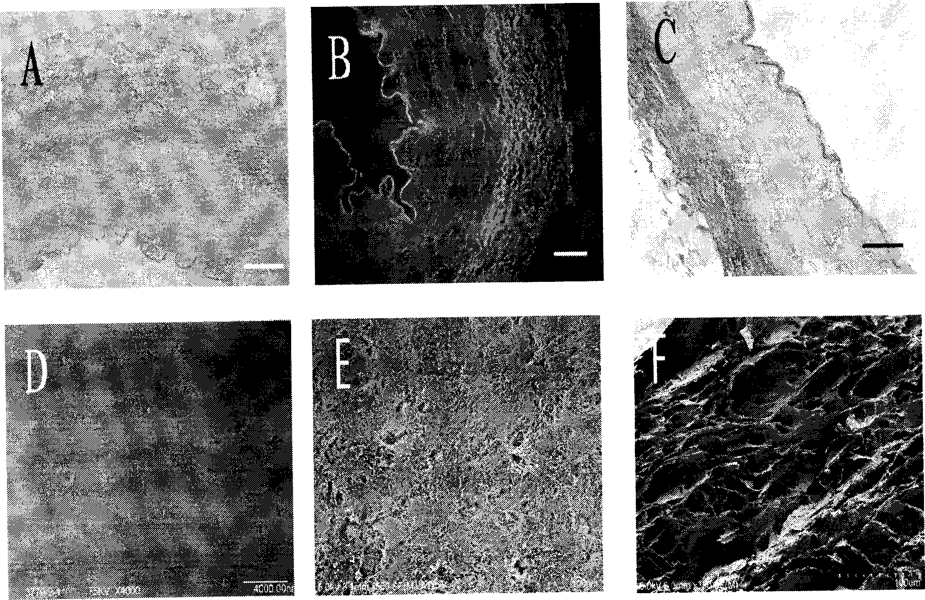
[0060] 1. Histology

[0061] Methods: Fix with 4% formaldehyde solution, observe the general structure and cell components of the acellular matrix by HE staining; observe the structural changes of collagen fibers and elastic fibers by Masson staining; observe the cell nucleus by DAPI fluorescent staining;

[0062] see results figure 1 A-1C.

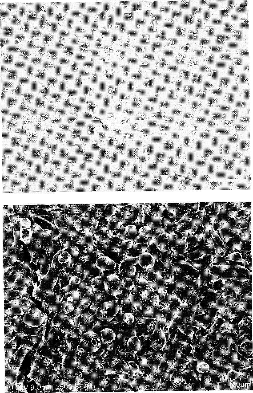
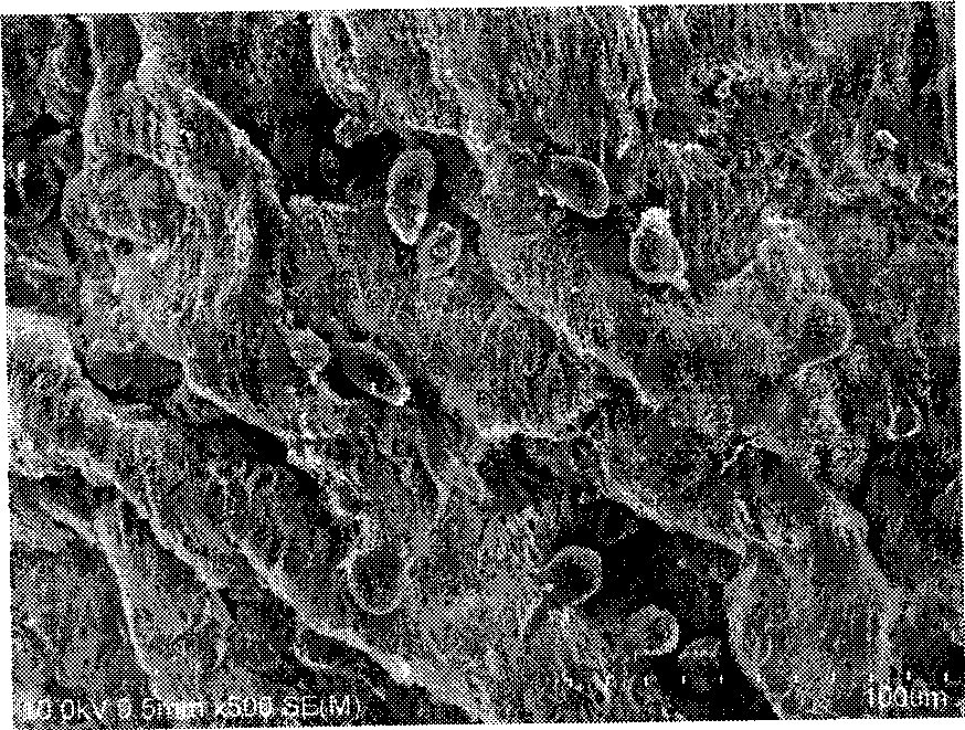
[0063] 2. Electron microscope observation

[0064] Methods: The cellular components and matrix components of the acellular matrix were observed by transmission electron microscope; the changes of tissue cell structure on the surface of the tube wall were observed by scanning electron microscope.

[0065] see results figure 1 D~1F. HE( figure 1 A) and DAPI ( figure 1 B) The staining results showed that the cellular components of the porcine iliac vessels were completely removed after being sequentially treated with 1% TritonX-100, 0.1% ammonia water and 0.25% trypsin-0.0...


PUM


Abstract

The invention relates to a blood vessel matrix for removing cell components in vascular tissue and a preparing method thereof. The invention provides a method which can remove the cell components in the vascular tissue and protect the structural integrity of the cell matrix at utmost. The method comprises the steps of cell extraction, detergent removing and enzyme treatment. The blood vessel cell removing matrix prepared by the invention has the advantages of excellent mechanical strength, proper porosity and low cytotoxicity and can successfully implant cells into the cell matrix, thereby providing a favorable tissue stent for the blood vessel tissue engineering.

Description

technical field [0001] The invention relates to a blood vessel matrix depleted of cells in blood vessel tissue and a preparation method thereof. Background technique [0002] In the treatment of cardiovascular diseases, arterial bypass surgery is the main method of vascular reconstruction. However, many patients cannot find suitable autologous blood vessels because of vascular diseases or vascular surgery. In the treatment of small-caliber vascular diseases, such as coronary arteries and sub-knee vessels, due to the lack of anticoagulated endothelial layer, or intimal hyperplasia caused by the mismatch between the compliance of artificial blood vessels and natural blood vessels, the long-term patency rate of artificial blood vessels is low. not ideal. [0003] After the animal blood vessels are decellularized, the cellular components that cause rejection in the vascular tissue are removed, and only the extracellular matrix components that are well conserved among animal sp...

Claims


Application Information

Patent Timeline
IPC IPC(8): A61L27/36
Inventor 董建德张建谷涌铨李春民李建新陈兵吴英锋
Owner XUANWU HOSPITAL OF CAPITAL UNIV OF MEDICAL SCI
Why Eureka
Try Eureka