Method for preparing Ge sealed radioactive source

A technology of radioactive source and purpose, applied in the field of medical devices, can solve the problems of poor radiation resistance, change in dose distribution, affecting imaging quality, etc., and achieve the effects of good uniformity, improved accuracy, and good adsorption effect.

Active Publication Date: 2009-12-02
天津赛德生物制药有限公司
View PDF0 Cites 14 Cited by
  • Summary
  • Abstract
  • Description
  • Claims
  • Application Information

AI Technical Summary

Problems solved by technology

However, this method is carried out under low-concentration acidic, neutral or alkaline conditions, so the radioactive germanium-68 solution needs to be converted into a low-acid solution during the preparation process. The uniformity of filling; most ion exchange resins have poor radiation resistance, and long-term irradiation will cause the resin to age or break, resulting in changes in dose distribution and affecting the quality of imaging

Method used


Image

  • Method for preparing Ge sealed radioactive source
  • Method for preparing Ge sealed radioactive source

Examples


Embodiment 1

[0019] 68 The preparation method of Ge sealed radioactive source is made up of the following steps:

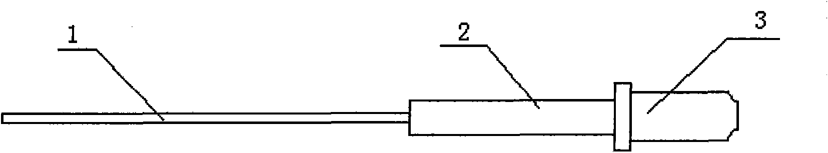
[0020] In a 15mL square quartz reaction bottle, put 2g of 120-mesh silicon dioxide as an adsorbent, add 5mL of sulfuric acid aqueous solution with a concentration of 10mol / L, and a radioactive concentration of 3mCi / mL 68 Ge aqueous solution 0.6mL, fix the reaction bottle on the rotary oscillator, under normal pressure, rotate and oscillate at a speed of 40 rpm for 60 minutes, take out the supernatant, wash the precipitate with deionized water for 3 times, and then use Wash it twice with acetone, dry it, put it into the stainless steel tube 1, weld the two ends, set the sealed stainless steel tube in the casing 2 and assemble it on the base 3, and then make a 68 Ge sealed radioactive source. The utilization rate of radioactive germanium-68 is 97%.

Embodiment 2

[0022] 68 The preparation method of Ge sealed radioactive source is made up of the following steps:

[0023] In a 15mL square quartz reaction bottle, put 2g of 80-mesh silicon dioxide as an adsorbent, add 5mL of sulfuric acid aqueous solution with a concentration of 15mol / L, and a radioactive concentration of 3mCi / mL 68 Ge aqueous solution 0.6mL, fix the reaction bottle on the rotary oscillator, under normal pressure, rotate and oscillate at a speed of 30 rpm for 120 minutes, take out the supernatant, wash the precipitate with deionized water for 3 times, and then use Clean it twice with ethanol, dry it, put it into a stainless steel tube, weld the two ends to seal, set the sealed stainless steel tube in the casing and assemble it on the base to make a 68 Ge sealed radioactive source. The utilization rate of radioactive germanium-68 is 95%.

Embodiment 3

[0025] 68 The preparation method of Ge sealed radioactive source is made up of the following steps:

[0026] In a 15mL square quartz reaction bottle, put 1.5g of 200-mesh silicon dioxide as an adsorbent, add 5mL of sulfuric acid aqueous solution with a concentration of 9mol / L, and a radioactive concentration of 3mCi / mL 68 Ge aqueous solution 0.6mL, fix the reaction bottle on a rotary oscillator, under normal pressure, rotate and oscillate at a speed of 45 rpm for 30 minutes, take out the supernatant, wash the precipitate 3 times with deionized water, and then use Clean it twice with ethanol, dry it, put it into a stainless steel tube, weld the two ends to seal, set the sealed stainless steel tube in the casing and assemble it on the base to make a 68 Ge sealed radioactive source. The utilization rate of radioactive germanium-68 is 96%.

[0027] The wall thickness of the stainless steel pipes in Examples 1-3 is 0.1 mm.


PUM


Abstract

The invention discloses a method for preparing a Ge sealed radioactive source, which consists of the following steps: putting 0.2 to 50 g of silicon dioxide with grain diameter of 50 to 400 meshes as an adsorbent in a reaction vessel, and adding 1 to 50 mL of 8 to 15 mol/L sulfuric acid water solution, 0.1 to 10 mL of Ge water solution with the radioactive concentration of 0.1 to 100 mCi/ mL and 0 to 15 mL of 0.1 to 50 g/L water solution of a carrier germanium; performing stirring or oscillatory reaction on the mixture for 10 to 120 minutes under normal pressure; taking supernatant out of the reaction vessel, and washing and drying the sediment to be loaded into a stainless steel tube; and performing solder sealing on two ends of the tube, arranging the sealed stainless steel tube in a bushing, and assembling the bushing on a pedestal to prepare the Ge sealed radioactive source. The method has simple and convenient preparation process, the adsorbed solution can be reused, the pollution to the environment is reduced, and the cost for treating radioactive waste is reduced.

Description

technical field [0001] The invention belongs to the technical field of medical devices, and in particular relates to a 68 Preparation method of Ge sealed radioactive source. Background technique [0002] Positron emission tomography (PET), also known as molecular imaging or biochemical imaging, is a new technology developed in nuclear medicine. Horizontal nuclear medicine diagnostic sign. It uses positron radionuclide-labeled drugs such as glucose, amino acids, nucleic acids, and ligands as tracers to display the metabolism, function, blood flow, cell proliferation, and receptors of cells in the body and lesion tissue at the molecular level in the form of anatomical images. Body distribution and other conditions can provide more physiological and pathological diagnostic information for clinic. PET has been widely used in clinical diagnosis of various diseases, therapeutic effect and basic medical research. Due to the inconsistency of the detector, the decay of the isotop...

Claims


Application Information

Patent Timeline
Patent Type & Authority Applications(China)
IPC IPC(8): G21G4/08A61B6/02
Inventor 阎尔坤
Owner 天津赛德生物制药有限公司
Why Eureka
Try Eureka