Multifunctional ultrasound contrast agent and preparation method thereof

An ultrasonic contrast agent and multi-functional technology, applied in the field of medicine, can solve the problems of increasing the metabolic burden of the body, side reactions of the contrast agent, etc., and achieve the effects of good imaging effect, enhanced ultrasonic imaging, and simple synthesis process.

Active Publication Date: 2011-01-26
CHONGQING MEDICAL UNIVERSITY
View PDF3 Cites 15 Cited by
  • Summary
  • Abstract
  • Description
  • Claims
  • Application Information

AI Technical Summary

Problems solved by technology

[0006] The present invention provides a multi-functional ultrasound contrast agent in order to realize "one probe with multiple functions" in view of the need to use different contrast agents for different imaging techniques, which not only increases the metabolic burden of the body, but also has side effects of contrast agents.

Method used


Image

  • Multifunctional ultrasound contrast agent and preparation method thereof
  • Multifunctional ultrasound contrast agent and preparation method thereof
  • Multifunctional ultrasound contrast agent and preparation method thereof

Examples


Embodiment 1

[0025] Example 1 Preparation of multifunctional contrast agent of the present invention

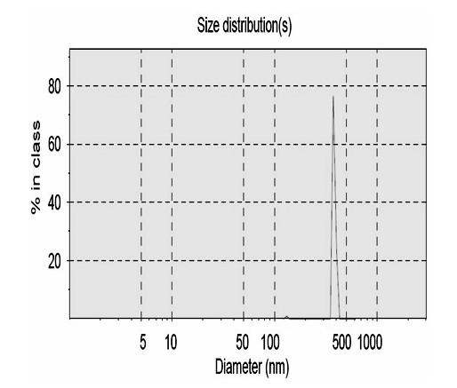
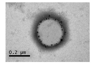
[0026] Precisely weigh 200 mg of lecithin and 40 mg of cholesterol and dissolve them together in 5 ml of chloroform solution. Rotate evaporate in a water bath at 55°C until a uniform film is formed. Add 8 ml of PBS buffer to make the film fall off and dissolve to obtain a lipid suspension. Liquid, the lipid suspension obtained above and 3 ml perfluorooctyl bromide were transferred to a high-speed disperser for emulsification, and 10 mg of Fe coated with lauric acid was added to the emulsification 3 o 4 Magnetic nanoparticles were emulsified for 10 min, then ultrasonically treated in an ice bath for 3 min, centrifuged (5 min, 3500 r / min) to remove the precipitate, and the upper liquid was collected to obtain Fe-loaded 3 o 4 Liquid fluorocarbon nanoparticles. Observation under a light microscope shows that the nanoparticles are spherical, small and uniform, regular in shape, and well d...

Embodiment 2

[0027] Example 2 In Vivo Ultrasonic Imaging Experiment of the Multifunctional Ultrasonic Contrast Agent of the Present Invention

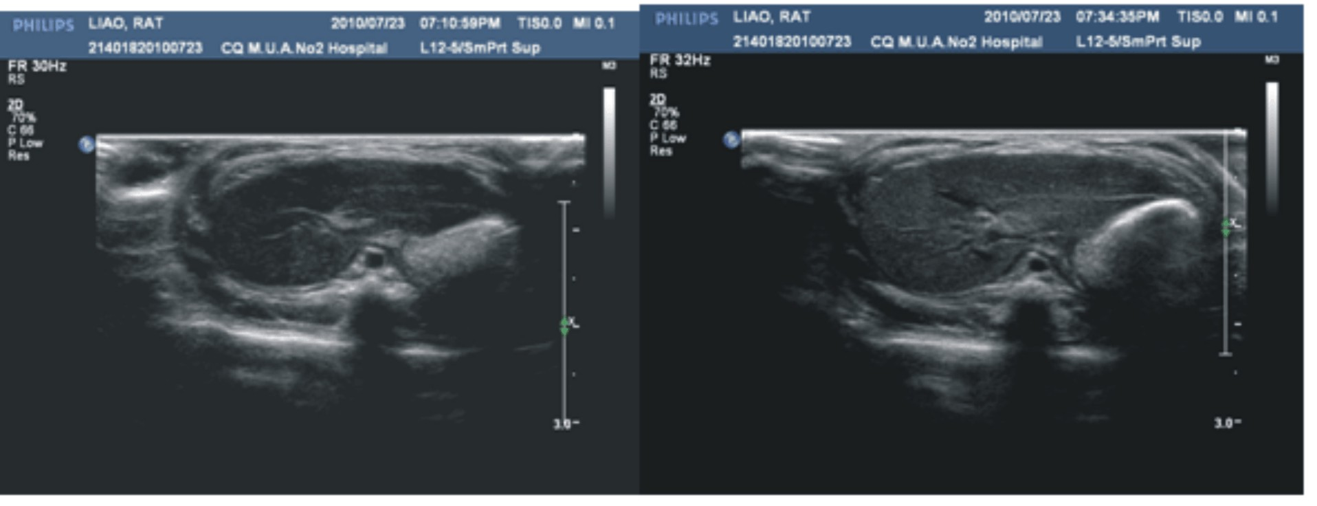
[0028] SD rats were anesthetized by intraperitoneal injection of 3% pentobarbital sodium (1 mg / kg), fixed in the supine position, and depilated. Using Philips iU22 color Doppler ultrasonic diagnostic instrument (L12-5 probe, probe frequency 7-13 MHz), using the method of self-control before and after, conventional scans were used to obtain rat liver sonograms, and a dose of 5 ml / kg was used for imaging. Rats were injected into the tail vein of the multifunctional ultrasound contrast agent prepared in Example 1, and the imaging time and imaging effect after hepatography were observed. Results Immediately after the injection of the contrast agent, there was no obvious enhancement of the liver parenchyma, but 5 minutes after the injection, the enhancement of the liver parenchyma began to appear, and reached the peak at about 20 minutes after the inj...

Embodiment 3

[0029] Example 3 In Vivo CT Imaging Experiment of the Multifunctional Ultrasound Contrast Agent of the Present Invention

[0030]Experimental animal preparation is the same as in Example 2. A GE LightSpeed ​​16-slice spiral CT machine was used to collect CT images of rat livers before contrast and 24 hours after contrast. CT scan conditions: tube voltage 100 kV, tube current 170 mA, slice thickness 5 mm. The results showed that the image density of rat liver parenchyma significantly increased 24 h after contrast, see Figure 4 . It shows that the multifunctional ultrasound contrast agent can enhance CT imaging. Different from the traditional CT iodine contrast agent, it is a contrast agent for blood pool imaging, which can be cleared quickly in the body. The contrast agent of the present invention has high stability in vivo and a long circulation time. Enhancement can still be seen after 24 hours, which is beneficial to the dynamic monitoring of lesions.


PUM


Abstract

The invention relates to a novel multifunctional ultrasound contrast agent and a preparation method thereof. The multifunctional ultrasound contrast agent of the invention is a nanoparticle lipid emulsion containing a core-shell structure, wherein materials of a shell membrane comprise lecithin, cholesterin and magnetic nanometer materials; the center is coated by lqiuid fluorocarbon; and magnetic nanoparticles are inlaid on the shell membrane. The ultrasound contrast agent of the invention is a specificity contrast agent of a reticuloendothelial system, thereby being capable of not only strengthening ultrasonoscopy, but also enhancing CT and MRI development, having good development effects, and having wide application prospects.

Description

technical field [0001] The invention relates to the field of medicine, in particular to a multifunctional novel ultrasonic contrast agent and a preparation method thereof. Background technique [0002] With the completion of human genome sequencing and the arrival of the post-genome era, the development of non-invasive and visualized imaging technology has attracted more and more attention and attention from the scientific community. In 1999, Weissleder et al. proposed the concept of molecular imaging, that is, the qualitative and quantitative research on biological processes at the cellular and molecular levels in vivo. This is a new discipline born from the combination of traditional medical imaging technology and molecular biology. Since the development of molecular imaging, it has been rated by the American Medical Association as one of the 10 frontier fields of medical science with the most development potential in the future, and is known as the medical imaging of the ...

Claims


Application Information

Patent Timeline
Patent Type & Authority Applications(China)
IPC IPC(8): A61K49/22A61K49/18A61K49/04A61K49/06
Inventor 王志刚冉海涛郑元义
Owner CHONGQING MEDICAL UNIVERSITY
Why Eureka
Try Eureka