Minimally invasive blood glucose monitoring microneedle and preparation method thereof

A blood sugar detection and microneedle technology, which is applied in diagnostic recording/measurement, medical science, sensors, etc., can solve problems such as interference, achieve the effect of maintaining enzyme activity, long-term preservation, and less damage

Inactive Publication Date: 2012-06-27
SOUTHEAST UNIV
View PDF3 Cites 24 Cited by
  • Summary
  • Abstract
  • Description
  • Claims
  • Application Information

AI Technical Summary

Problems solved by technology

However, analyte-like biomolecules can interfere with ...

Method used


Image

  • Minimally invasive blood glucose monitoring microneedle and preparation method thereof
  • Minimally invasive blood glucose monitoring microneedle and preparation method thereof
  • Minimally invasive blood glucose monitoring microneedle and preparation method thereof

Examples


Embodiment 1

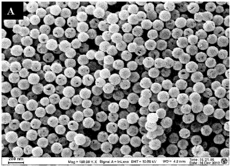
[0040] Embodiment 1: the preparation of a kind of gold nanoparticle

[0041] Preparation of gold nanoparticles The method of reducing chloroauric acid with sodium borohydride was used to prepare gold nanoparticles with a diameter of 50 nm. Add 3ml of 1% chloroauric acid solution into 200ml of stirred pure water, and then add 1ml of 0.2M potassium carbonate solution. Finally, 3 ml of freshly prepared 0.5 mg / ml sodium borohydride solution was quickly added. The solution turned from bright yellow to purple-black and then to wine red. Continue to stir for 5 minutes, and store in a 4°C refrigerator for later use.

Embodiment 2

[0042] Embodiment 2: the preparation of a kind of silver nanoparticles

[0043] Preparation of silver nanoparticles The method of reducing silver nitrate with sodium citrate was used to prepare silver nanoparticles with a diameter of 40 nm. Take 3ml of 1% silver nitrate solution and add it into 200ml of pure water stirred, then add 5ml of 0.1M sodium citrate solution and heat to boiling, then heat for 5 minutes and then cool naturally, the solution turns colorless and gradually turns bright yellow. After reaching room temperature, store in a 4°C refrigerator for later use.

Embodiment 3

[0044] Example 3: Preparation of a gold shell silica core particle

[0045] Gold nanoshell particles were prepared with spherical silica particles with a diameter of 110 nm as the core. Add 5 microliters of 3-aminoethoxysilane to 100 milliliters of alcohol sol containing 1 gram of silica, overnight at 37°C. Then transfer to an oven (80° C.) for curing for 3 hours. Centrifuge to remove excess 3-aminoethoxysilane to obtain aminated silica. The aminated silicon dioxide was added into the gold nanoparticle colloidal solution and stirred for 3 hours, and the silicon dioxide / gold nanocomposite particles were separated by centrifugation. The silicon dioxide / gold nanocomposite particles are added to 0.01% chloroauric acid solution, and then reducing agents such as formaldehyde or hydrogen peroxide are added to deposit the chloroauric acid on the surface of the silicon dioxide to form gold nanoshells.


PUM

PropertyMeasurementUnit
Particle sizeaaaaaaaaaa

Abstract

The invention discloses a minimally invasive blood glucose monitoring microneedle which comprises a needle handle, a needle body and a needle point which are sequentially connected, wherein after one of an amino group, an aldehyde group, a hydroxide radical and a carboxyl group is modified on the surfaces of the needle body and needle point of the microneedle, and the microneedle can be covered by a metal nano material layer modified by a Ph response molecule, the microneedle is inserted in a solution consisting a water-soluble polymer and a water-insoluble polymer, a porous polymer material layer is coated on the metal nano material layer, and then the microneedle is inserted in a glucose oxidase solution, so that the water-soluble polymer is dissolved and a plurality of pores are formed in a polymer layer; and glucose oxidase is absorbed on the surface of the porous polymer layer or in the holes, and the metal nano material has a grain size of 20-1000nm. By using the characteristic that the minimally invasive blood glucose monitoring microneedle has different intensities of Raman signals for different concentrations of glucose, the purpose of minimally invasively detecting the glucose in vivo and in vitro can be achieved.

Description

technical field [0001] The invention relates to the technical field of minimally invasive blood sugar detection, more specifically, a minimally invasive blood sugar detection microneedle with enhanced Raman signal and a preparation method thereof. Background technique [0002] Diabetes mellitus (Type I and Type II) has become an important health-threatening disease and tends to be younger. The cost of diagnosing, monitoring and treating diabetes is increasing year by year. Wide fluctuations in blood sugar levels in people with diabetes can lead to a variety of complications, including kidney disease, heart disease, blindness, nerve damage, and gangrene. For different types of diabetes, the current treatment methods are mainly through frequent monitoring of blood sugar levels, combined with appropriate diet, medication and insulin injections. Most patients use a fingerstick device to obtain a small blood sample to measure blood glucose levels, and then monitor glucose by in...

Claims


Application Information

Patent Timeline
IPC IPC(8): A61B5/1495
Inventor 董健陶琴许蓓蓓钱卫平
Owner SOUTHEAST UNIV
Why Eureka
Try Eureka