Medical nano-fiber sponge material and preparation method and application thereof

A nanofiber, sponge technology

Active Publication Date: 2014-05-07
ZHEJIANG UNIV
View PDF4 Cites 0 Cited by
  • Summary
  • Abstract
  • Description
  • Claims
  • Application Information

AI Technical Summary

Problems solved by technology

However, conventional pasty hydrogel dressings have poor air permeability. Although the freeze-dried sponge dressing has improved air permeability, the pore wall of the ice crystal particle-induced sponge dressing is a continuous sheet structure, and the permeability and microstructure are low.
Technologies such as high-voltage electrospinning, self-assembly, and phase separation have solved the problem of nanofiber structure formation, but chemical crosslinking has greatly reduced the anti-blocking properties of the material (Sung HW, et al. J Biomed Mater Res . 1999, 46 , 520)

Method used


Image

  • Medical nano-fiber sponge material and preparation method and application thereof
  • Medical nano-fiber sponge material and preparation method and application thereof
  • Medical nano-fiber sponge material and preparation method and application thereof

Examples


Embodiment 1

[0037] 1) NaNO 3 and Ca(NO 3 ) 2 4H 2 O was dissolved in 20 mL deionized water at a molar ratio of 1:0.5, NaNO 3 The concentration of the solution is 0.6 mol / liter, adjust the pH value to 12 with ammonia water, stir continuously, and set aside;

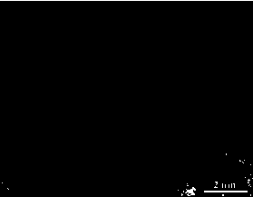
[0038] 2) Add triethyl phosphate and ethyl orthosilicate to 80 mL of absolute ethanol at a molar ratio of 1:25, and the molar concentration of ethyl orthosilicate is 0.5 mol / L. After stirring evenly, add to the above step 1 ) in the inorganic salt mixed solution, stirring continuously for 60 minutes, at 60 o C aged for 72 hours, then dried, and at 550 o Calcined at C to obtain bioactive glass particles, such as figure 1 As shown, the particle size of the particles is 200-300 nm.

[0039] 3) Add 0.1 g of bioactive glass particles obtained in step 2) to 20 ml of gelatin hydrosol with a mass percentage of 10%, stir and mix well, add 25 ml of absolute ethanol to it, continue stirring evenly, and place in -80 o Stand still in C l...

Embodiment 2

[0046] 1) NaNO 3 and Ca(NO 3 ) 2 4H 2 O was dissolved in 20 mL deionized water at a molar ratio of 1:0.8, NaNO 3 The concentration of the solution is 0.5 mol / liter, adjust the pH value to 11 with ammonia water, stir continuously, and set aside;

[0047] 2) Add triethyl phosphate and ethyl orthosilicate to 80 mL of absolute ethanol at a molar ratio of 1:30, and the molar concentration of ethyl orthosilicate is 0.5 mol / L. After stirring evenly, add to the above step 1 ) in the inorganic salt mixed solution, stirring continuously for 90 minutes, at 80 o C aged for 48 hours, then dried, and at 520 o C and calcined to obtain bioactive glass particles with a particle size of 270-380 nm.

[0048] 3) Add 0.15 g of bioactive glass particles obtained in step 2) to 25 ml of gelatin hydrosol with a mass percentage of 10%, stir and mix well, add 30 ml of absolute ethanol to it, continue to stir evenly, and place in- 80 o Stand still in C low temperature equipment for 20 hours to ob...

Embodiment 3

[0054] 1) NaNO 3 and Ca(NO 3 ) 2 4H 2 O was dissolved in 20 mL deionized water at a molar ratio of 1:1.2, NaNO 3 The concentration of the solution is 0.3 mol / liter, adjust the pH value to 11 with ammonia water, stir continuously, and set aside;

[0055] 2) Add triethyl phosphate and ethyl orthosilicate to 80 mL of absolute ethanol at a molar ratio of 1:20, and the molar concentration of ethyl orthosilicate is 0.5 mol / L. After stirring evenly, add to the above step 1 ) in a mixed solution of inorganic salts, stirring continuously for 90 minutes, at 75 o C aged for 60 hours, then dried, and at 600 o C and calcined to obtain bioactive glass particles with a particle size of 430-620 nm.

[0056] 3) Add 0.18 g of bioactive glass particles obtained in step 2) to 30 ml of gelatin hydrosol with a mass percentage of 8%, stir and mix well, add 25 ml of absolute ethanol to it, continue stirring evenly, and place in -80 o Stand still in C low temperature equipment for 20 hours to ...


PUM

PropertyMeasurementUnit
diameteraaaaaaaaaa
particle sizeaaaaaaaaaa

Abstract

The invention relates to a medical nano-fiber sponge material, of which the porosity is 90-98 percent and the diameter of a nano-fiber is 50-1,000 nanometers. The medical nano-fiber sponge material consists of the following components in percentage by mass: 0.5-8.5 percent of bioactive glass fine particles, 65-88 percent of gelatin, 0.2-5.0 percent of hyaluronic acid, 0.2-5.0 percent of chitosan and the balance of water. A preparation method of the medical nano-fiber sponge material comprises the following steps of: adding bioactive glass fine particles and ethanol into a gelatin aqueous solution, and stirring uniformly; performing low-temperature phase separation and freeze drying to obtain nano-fiber sponge obtained by compounding gelatin and bioactive glass fine particles; and performing soaking, refrigerating and freeze drying with a hyaluronic acid solution and a chitosan solution in sequence under a negative pressure condition to obtain a porous composite sponge material in which nano-fibers are crosslinked and coated statically with hyaluronic acid and chitosan. The nano-fiber sponge has excellent bioactivity, bacteriostasis and mechanical property, and can be widely applied in the fields of regenerative repair of various skin wounds and skin tissue engineering.

Description

technical field [0001] The invention relates to a medical nanofiber sponge material and its preparation method and application, belonging to the fields of biomedical materials and wound repair. Background technique [0002] In the 1960s, Winter proposed the theory of "moist airtight therapy". He found that the epidermis of non-scab wounds healed faster in a moist airtight environment than in dry and scab-like conditions. Therefore it is recognized that the purpose of using dressings is not only to prevent the invasion of germs, dressings also need to help and promote wound healing. At present, clinical temporary wound dressings are divided into biological dressings, synthetic dressings and biosynthetic dressings according to the source of materials. Biological dressings are mainly film and sponge dressings prepared by reprocessing biological tissues such as pigskin, amniotic membrane, and bovine collagen. They can prevent excessive evaporation of water, effectively iso...

Claims


Application Information

Patent Timeline
Patent Type & Authority Patents(China)
IPC IPC(8): A61L15/32A61L15/28A61L15/18C08J9/42
Inventor 苟中入杨贤燕陈晓怡
Owner ZHEJIANG UNIV
Why Eureka
Try Eureka