Method and chip for detecting tumor cell EGFR (epidermal growth factor receptor) gene mutation

A tumor cell and chip technology, which is applied in biochemical equipment and methods, microbial determination/examination, chemical libraries, etc., can solve the problems of time-consuming, labor-intensive, high cost, and complicated operation

Inactive Publication Date: 2012-10-17
HAINAN MEDICAL COLLEGE
View PDF5 Cites 11 Cited by
  • Summary
  • Abstract
  • Description
  • Claims
  • Application Information

AI Technical Summary

Problems solved by technology

In 2004, Lynoh and Paeg first discovered that EGFR gene mutations are related to the sensitivity of gefitinib and other drugs. The total effective rate of gefitinib in the treatment of NSCLC (non-small cell lung cancer) is 10-30%. 75-95%, the effective rate of tumors without gene m...

Method used


Image

  • Method and chip for detecting tumor cell EGFR (epidermal growth factor receptor) gene mutation
  • Method and chip for detecting tumor cell EGFR (epidermal growth factor receptor) gene mutation
  • Method and chip for detecting tumor cell EGFR (epidermal growth factor receptor) gene mutation

Examples


Embodiment 1

[0064] Embodiment 1: chip preparation

[0065] Materials and reagents: Aldehyde glass substrate, gene microarray hybridization box, chip cover slip, chip fence, microarray chip centrifuge tube (purchased from Beijing Boao Biotechnology Company); chip spotting instrument (SpotBot Personal Microarray Robot) ) are products of Telechem Corporation.

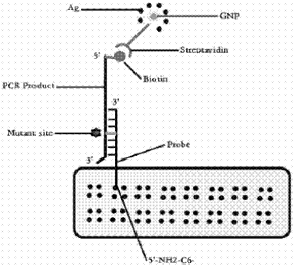
[0066] Oligonucleotide probe design and synthesis: use OLIGO6.0 software to design oligonucleotide probes (see SEQ ID NO:9-24), 5'-terminal amino modification (5'-NH 2 -(CH 2 ) 6 -), the specificity of all probes was confirmed by BLAST comparative analysis, and the probes were synthesized and purified by HPLC by Yingjun Biotechnology Company.

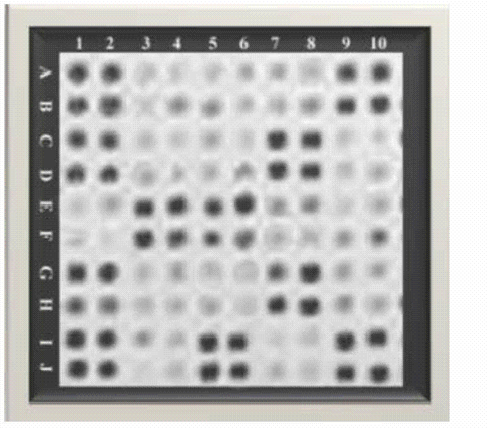
[0067] Chip preparation: 5'-NH 2 -(CH 2 ) 6 -Modify each probe, dilute to 20 μmol / L with spotting solution, use SpotBot Personal Microarray Robot automatic spotting instrument, SMP10B spotting needle to load the probe on the aldehyde glass slide, spotting diameter 365 μm, spacing 460 μm, ...

Embodiment 2

[0068] Embodiment 2: Tumor cell DNA extraction and PCR amplification

[0069] Sample to be tested: paraffin-embedded formalin-fixed (FFPE) tumor tissue;

[0070] First deparaffinize, put 5-8 slices of 5-10μm thick FFPE tumor tissue into a 1.5ml centrifuge tube, incubate at 72°C for 20min, remove the dissolved paraffin liquid with a pipette, add 1ml xylene preheated to 50°C Solution, incubated at 50°C for 10 minutes, centrifuged at 12000g for 2 minutes, and then sucked off the xylene with a pipette. Repeat the operation of heating and removing the paraffin liquid 2 times. Add 1ml of absolute ethanol and place it at room temperature for 10min, centrifuge at 12000g for 5min, absorb the ethanol with a pipette, then add 90% and 70% ethanol in turn to repeat the hydration treatment, and finally wash and dry with deionized water. DNA was extracted with a paraffin-embedded tissue DNA extraction kit (QIAamp DNA FFPE tissue kit, QIAGEN) and operated according to the instructions. The...

Embodiment 3

[0072] Example 3: Preparation of streptavidin-coupled gold nanoparticles (Streptavidin-AuNP)

[0073] The pH value of the gold nanoparticle solution was adjusted to 6.5 with hydrochloric acid, and 100 μl (250 ng / μl) streptavidin was added to every 100 μl AuNP solution, and gently shaken at room temperature for 5 min. Add 100μl 10% NaCl solution and incubate at room temperature for 2h. Add 5% BSA to a final concentration of 2%, incubate at room temperature for 15 min, centrifuge at 20,000 rpm for 45 min at 4°C, and discard the supernatant. Sodium azide buffer (20mM Tirs-HCl pH6.2, 2%BSA, 0.02%NaN 3 ) to wash the red precipitate, centrifuge at 20,000 rpm for 45 min at 4°C, and discard the supernatant. Repeat washing and centrifugation 3 times, then resuspend the pellet with sodium azide buffer, and store at 4°C for future use.


PUM


Abstract

The invention relates to the technical field of biology, and discloses a method for detecting tumor cell EGFR (epidermal growth factor receptor) gene mutation. The method comprises the following steps: taking sequences shown in SEQ ID NO:1-8 as primers and DNA (deoxyribonucleic acid) of a sample to be detected as a template to carry out PCR (polymerase chain reaction) amplification; and respectively hybridizing probes having sequences shown in SEQ ID NO:9-24 with the obtained amplification products, then washing to remove the uncombined amplification products, finally incubating hybrid products with streptavidin coupled with gold nanoparticles, and judging the mutation result according to color variations of the gold nanoparticles. Based on the chip technology, by utilizing the characteristic of specific binding of biotins and streptavidin, the streptavidin coupled with gold nanoparticles is specifically bound with the biotins labelled on the amplification products after the probes aiming at EGFR gene mutation related to sensitivity or drug resistance to gefinitib, erlotinib and the like compose a chip array and are hybridized with the biotin labelled amplification products, thus accurately interpreting the mutation result.

Description

technical field [0001] The invention relates to the field of biotechnology, in particular to a method and a chip for detecting tumor cell EGFR gene mutation. Background technique [0002] Epidermal growth factor receptor (EGFR), a member of the receptor tyrosine kinase superfamily, activates the serine / threonine protein kinase AKT through PI3K, activates the apoptosis inhibitory protein Caspase9, and inhibits apoptosis-promoting cells proliferation. [0003] Increased or persistent activation of EGFR will promote excessive cell proliferation and tumorigenesis, and is closely related to tumor cell behaviors such as tumor invasion and metastasis, angiogenesis, and anti-apoptosis. Studies have found that the expression of EGFR is increased in 40-80% of lung cancer, 14-91% of breast cancer, 33-74% of gastric cancer, 40-80% of prostate cancer and 36-100% of head and neck tumors, and its expression The level of EGFR is negatively correlated with the degree of malignancy, and the...

Claims


Application Information

Patent Timeline
IPC IPC(8): C12Q1/68C40B40/06
Inventor 白玉杰薛丽
Owner HAINAN MEDICAL COLLEGE
Why Eureka
Try Eureka