Cultural method for inducing human embryonic stem cell to directionally differentiate into corneal limbal stem cell

A technology of corneal limbal stem cells and human embryonic stem cells, which is applied in the direction of non-embryonic pluripotent stem cells, artificially induced pluripotent cells, animal cells, etc., can solve the problems of low induction efficiency and inability to meet the clinical treatment of ocular surface diseases, and achieve reduction The effect of scar formation, repair of damaged ocular surface, and reduction of inflammatory response

Active Publication Date: 2013-03-06
SHANDONG UNIV
View PDF1 Cites 16 Cited by
  • Summary
  • Abstract
  • Description
  • Claims
  • Application Information

AI Technical Summary

Problems solved by technology

However, the existing embryonic stem cell induction and differentiation schemes have low induction efficiency, and the induced cells are all terminally differentiated corneal epithelial cells, which do not have the phenotype of normal limbal stem cel

Method used


Image

  • Cultural method for inducing human embryonic stem cell to directionally differentiate into corneal limbal stem cell
  • Cultural method for inducing human embryonic stem cell to directionally differentiate into corneal limbal stem cell
  • Cultural method for inducing human embryonic stem cell to directionally differentiate into corneal limbal stem cell

Examples


Embodiment 1

[0025] Example 1 Inducing Human Embryonic Stem Cells to Differentiate into Limbal Stem Cells and Preparation of Corneal Grafts

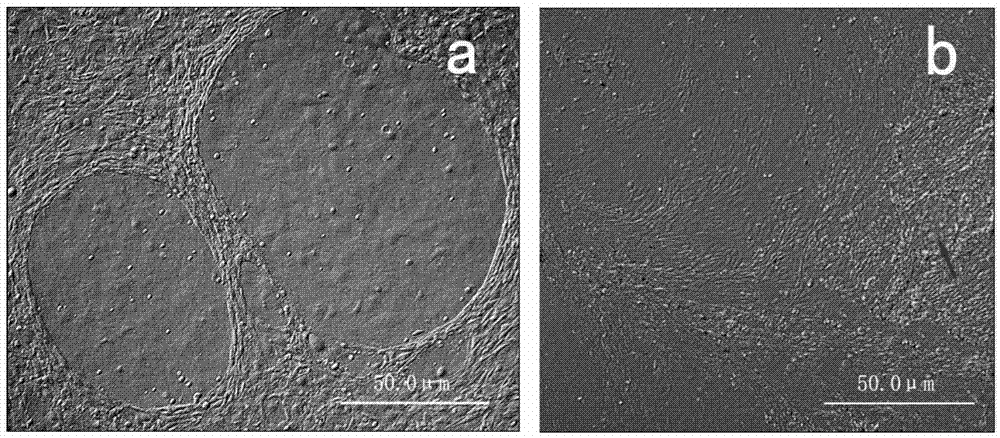
[0026] The fresh human amniotic membrane was rinsed and disinfected with PBS balanced salt solution containing 100 U / ml penicillin-streptomycin, digested with 0.25% trypsin at 37°C for 30 minutes, and the amniotic membrane cells were removed by pipetting to obtain the decellularized amniotic membrane scaffold material. The acellular amniotic membrane scaffold material was cut into discs with a diameter of 3 cm using ophthalmic scissors under a dissecting microscope. Human embryonic stem cells were cultured in induction medium for 9 days to differentiate into limbal stem cell-like cells (such as figure 1 b) Digest induced cells with 0.125% trypsin at 37°C for 5 minutes to prepare single cell suspension, press 1.5X10 4 cells / cm 2 Inoculate it on the cut acellular amniotic membrane scaffold material (the inoculation area of ​​the scaffold material is ab...

Embodiment 2

[0032] The tissue-engineered ocular surface constructed in Example 2 repairs chemically damaged corneal epithelium

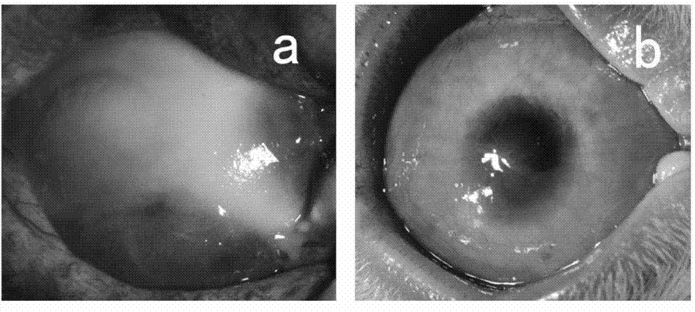
[0033] Soak the ring-shaped filter paper in 1mol / L soda ash for 20 seconds, absorb the excess liquid with sterile gauze, place it on the ocular surface of the rabbit and cauterize it for 40 seconds, then rinse the ocular surface with a large amount of sterile saline to prepare rabbit limbal stem cell degeneration compensation model. One month after the burn, select the successfully constructed rabbit corneal limbal stem cell decompensation model, carefully remove the new blood vessel fibrous membrane under the microscope, clean the implant bed, and place the induced cell graft (prepared in Example 1) on the prepared implant bed. Suture the edges of the graft with 10-0 nylon sutures. The implants were observed under the slit lamp every 3 days after the operation, and the cell growth was observed under the confocal microscope every 1 month. Three months after tr...

Embodiment 3

[0034] The tissue-engineered ocular surface constructed in Example 3 repairs mechanically damaged corneal epithelium

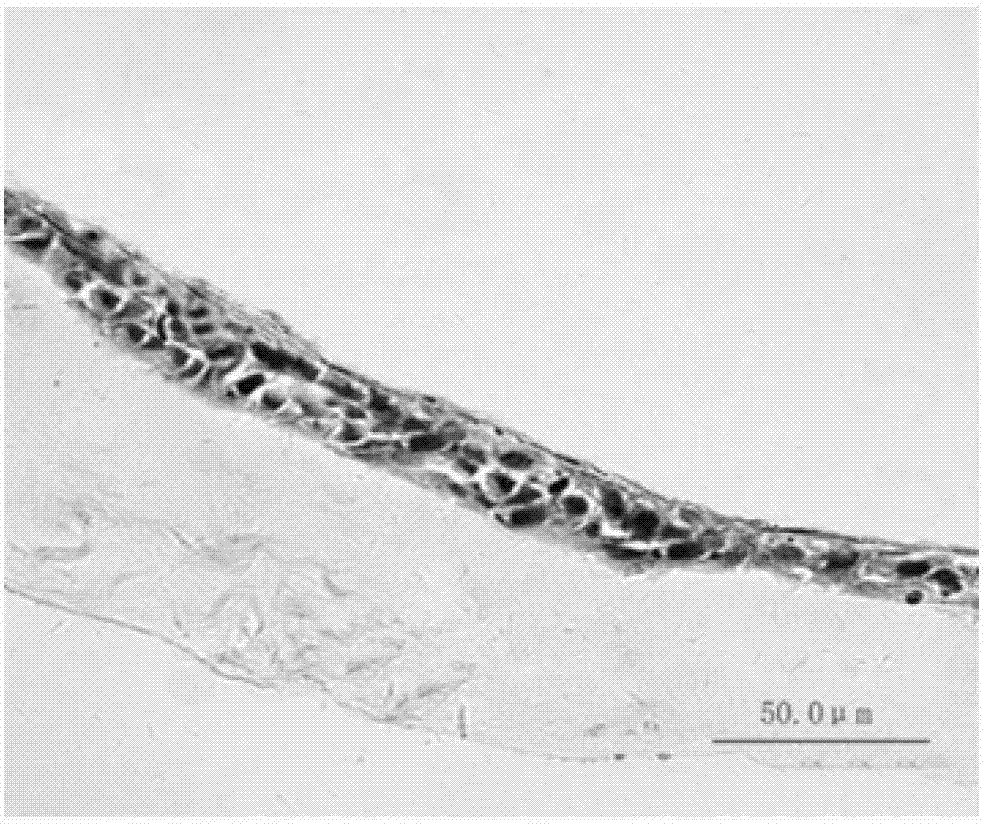
[0035] The rabbit corneal limbal tissue was excised circularly under a microscope with a lamellar knife to prepare a rabbit corneal limbal stem cell decompensation model. One month after the surgical injury, select the successfully constructed rabbit corneal limbal stem cell decompensation model, carefully remove the new blood vessel fibrous membrane under the microscope, clean the implant bed, and place the induced cell graft (prepared in Example 1) on the prepared implant bed , with 10-0 nylon suture at the edge of the graft. The implants were observed under the slit lamp every 3 days after the operation, and the cell growth was observed under the confocal microscope every 1 month. Three months after transplantation, the experimental animals were sacrificed for histological detection, such as Figure 4 shown.


PUM


Abstract

The invention discloses a cultural method for inducing a human embryonic stem cell to directionally differentiate into a corneal limbal stem cell, which comprises the following steps that firstly, a DMEM (Dulbecco's Modified Eagle Medium)/F12 conditioned medium is adopted to culture a human primary corneal limbal stem cell to prepare a corneal limbal stem cell conditioned medium, and then the corneal limbal stem cell conditioned medium is utilized and combined with IV type collagen culture in vitro to induce the human embryonic stem cell to directionally differentiate into the corneal limbal stem cell. According to the corneal limbal stem cell obtained by utilizing the cultural method, through light microscope observation in vitro, electron microscope observation, real-time quantitative polymerase chain reaction, immunofluorescence, flow cytometry, cloning efficiency determination and the like, the induced cell has a similar shape and phenotype with a normal corneal limbal stem cell, has good differentiation and proliferation capacity in vitro, can be transferred in vitro for more than four generations and can be used as a seed cell for preparing a corneal graft.

Description

technical field [0001] The invention relates to a culture method for inducing directional differentiation of human embryonic stem cells into limbal stem cells, and a method for constructing tissue-engineered ocular surface grafts using limbal stem cells obtained by the culture method combined with decellularized amniotic membrane scaffolds, belonging to the field of tissue engineering and ophthalmology . Background technique [0002] Limbal stem cells are the fundamental basis for the self-renewal, repair and regeneration of corneal epithelial cells, and are also an important barrier structure between the cornea and conjunctiva. They play an extremely important role in maintaining the transparency, stability and normal physiological functions of the cornea. Severe ocular surface diseases, such as ocular surface chemical injury, inflammation, ocular surface immune disease, etc., can lead to the loss of corneal limbal stem cells and the destruction of the barrier structure, th...

Claims


Application Information

Patent Timeline
IPC IPC(8): C12N5/074A61L27/38
Inventor 吴欣怡朱婧杨洪玲
Owner SHANDONG UNIV
Why Eureka
Try Eureka