Method for eliminating alpha-synuclein generated in Parkinson's model cells by using cadmium telluride quantum dots and application thereof

A technology of cadmium telluride quantum dots and nuclear synapse proteins, applied in animal cells, tumor/cancer cells, vertebrate cells, etc.

Active Publication Date: 2013-12-18
SHANGHAI INST OF APPLIED PHYSICS - CHINESE ACAD OF SCI
View PDF1 Cites 0 Cited by
  • Summary
  • Abstract
  • Description
  • Claims
  • Application Information

AI Technical Summary

Problems solved by technology

However, until now, there has been no research on the removal of toxic α-nuclear synapsin by effectively triggering autophagy in Parkinsonian model cells

Method used


Image

  • Method for eliminating alpha-synuclein generated in Parkinson's model cells by using cadmium telluride quantum dots and application thereof
  • Method for eliminating alpha-synuclein generated in Parkinson's model cells by using cadmium telluride quantum dots and application thereof
  • Method for eliminating alpha-synuclein generated in Parkinson's model cells by using cadmium telluride quantum dots and application thereof

Examples


Embodiment 1

[0023] Example 1 Preparation of water-soluble cadmium telluride quantum dots (prepared with reference to CN1693206A)

[0024] 1) Preparation of potassium hydride telluride: 110.5 mg KBH 4 Put the solid and 91.6 mg of tellurium powder into a flask, add 3 milliliters of water, and react at 0 degrees Celsius for 20 hours to obtain a KHTe solution for subsequent use;

[0025] 2) Preparation of cadmium telluride precursor solution: 36.8 mg of CdCl 2 Dissolve in 100 ml of water, add 0.05 ml of mercaptoacetic acid, adjust pH=11.0 with 0.5 mol / L NaOH solution to obtain cadmium telluride precursor solution, pass nitrogen to remove oxygen, and store under nitrogen protection;

[0026] 3) Preparation of CdTe quantum dots by program-controlled microwave irradiation: the prepared cadmium telluride precursor solution was subjected to program-controlled microwave heating to obtain water-soluble cadmium telluride quantum dots. The microwave heating conditions are as follows: the microwave p...

Embodiment 2

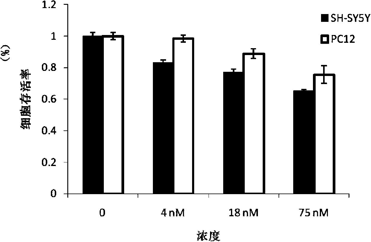
[0028] Example 2 Detection of biocompatibility of cadmium telluride quantum dots using thiazole blue method

[0029] Experimental method: Thiazolium blue method (MTT) is a method to detect cell survival and growth. The detection principle is that succinate dehydrogenase in the mitochondria of living cells can reduce exogenous thiazolyl blue to water-insoluble blue-purple crystalline formazan and deposit in the cells, while dead cells have no such function. Sodium dodecyl sulfate at pH 2.5 can dissolve formazan in cells, and its light absorption value is measured at a wavelength of 490nm with an enzyme-linked immunosorbent detector, which can indirectly reflect the activity of living cells.

[0030] The detection process includes the following steps:

[0031] 1) Cell culture: SH-SY5Y cells (derived from human dopaminergic neuroblastoma cell line, purchased from the Cell Bank of the Chinese Academy of Sciences) or PC12 cells (derived from rat adrenal medulla pheochromocytoma, p...

Embodiment 3

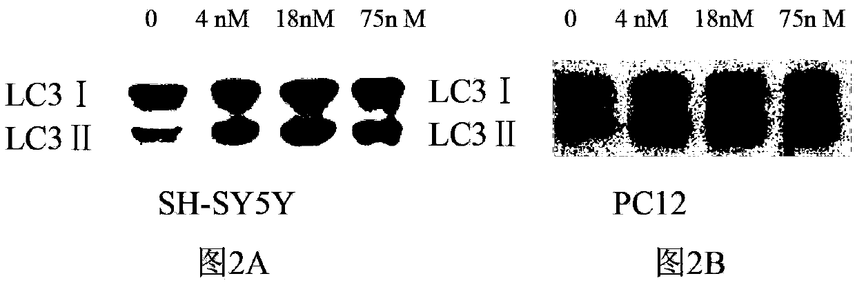
[0038] Example 3 Detection of whether autophagy occurs in cells

[0039] Experimental method: When cells undergo autophagy, LC3Ⅰ protein with a molecular weight of 18Kd in the cytoplasm combines with phosphatidylethanolamine on the surface of the autophagic vesicle membrane to form 16Kd LC3Ⅱ, which is located in the autophagosome membrane. The ratio of LC3II to LC3I protein content reflects the autophagy activity of cells. The present invention uses Western blot method to detect the value of LC3II / LC3I so as to detect the influence of quantum dots on cell autophagy.

[0040] The detection process includes the following steps:

[0041] 1) Cell culture: place SH-SY5Y or PC12 cells at 37°C with 5% CO 2 Culture in the DMEM medium that contains the fetal calf serum of volume fraction 10% and 1% penicillin streptomycin in the incubator;

[0042] 2) Cell plating: Digest the cell suspension after the cells are full, and count the cells at 3×10 cells per well. 5 Cells were plated in...


PUM

PropertyMeasurementUnit
particle diameteraaaaaaaaaa

Abstract

The invention relates to a method for eliminating alpha-synuclein generated in Parkinson's model cells by using cadmium telluride quantum dots and application thereof. The method comprises the following steps: spreading a cell suspension obtained by Parkinson's model cell digestion into 6 pore plates, wherein 3*10<4>-3*10<5> cells are spread in each hole; and after the cells are attached to walls, removing the culture medium in the hole, and adding a culture medium containing 4-75nM cadmium telluride quantum dots, thereby obviously eliminating the overexpressed alpha-synuclein in the Parkinson's model cells under the action of the cadmium telluride quantum dots. The Parkinson's model cells are SH-SY5Y cells which are constructed in vitro from 1-methyl-4-phenyl-pyridine ions and subjected to induced differentiation with all-trans-vitamin A acid and tetradecanoyl phorbol acetate, or differentiated PC12 cells constructed in vitro from 1-methyl-4-phenyl-pyridine ions. Cadmium telluride quantum dots with favorable biocompatibility are utilized to induce autophagy effect of cells, thereby eliminating overexpressed toxic protein (alpha-synuclein) in the Parkinson's model cell.

Description

technical field [0001] The invention belongs to the field of bionano materials, and in particular relates to a method for removing α-nuclear synapse protein produced in Parkinson's model cells by using cadmium telluride quantum dots. Background technique [0002] Parkinson's disease (PD) is the most common neurodegenerative disorder, affecting more than 0.1% of the world's population over the age of 40. Clinically, most patients show involuntary trembling, irritability, depression or mental abnormality when they are inactive and resting, which brings a great economic and mental burden to the society and the patient's family, so the research on this disease is becoming more and more serious. are getting more and more attention. The pathological manifestations of Parkinson's disease are the loss of dopamine neurons in the substantia nigra pars compacta and the increase of Lewy inclusion bodies. Lewy bodies are mainly composed of amyloid protein fibers called α-nuclear synaps...

Claims


Application Information

Patent Timeline
Patent Type & Authority Patents(China)
IPC IPC(8): C12N5/09
Inventor 黄庆陈楠李晓明魏敏樊春海
Owner SHANGHAI INST OF APPLIED PHYSICS - CHINESE ACAD OF SCI
Why Eureka
Try Eureka