
Preparation method and application of autologous mesenchymal stem cell-loaded human amniotic membrane cornea paster

A bone marrow mesenchymal and stem cell technology, applied in the field of regenerative medicine, can solve the problems of less treatment opportunities, increased risk of disease in healthy eyes, and limited source of materials

Inactive Publication Date: 2013-04-24
北京清美联创干细胞科技有限公司
View PDF5 Cites 9 Cited by
  • Summary
  • Abstract
  • Description
  • Claims
  • Application Information

AI Technical Summary

Problems solved by technology

[0004] 1. The short-term corneal transparency after traditional keratoplasty, its main disadvantage is that only the central φ7mm optically transparent area is transplanted. Due to the obvious rejection, long-term use of immunosuppressants is required to cooperate with the treatment
[0005] 2. Although there is no immune rejection in autologous corneal limbus transplantation, the source of materials is limited. The main disadvantage is that it increases the risk of disease in healthy eyes. It cannot be transplanted in a 360° circle, and the effect is limited.
[0006] 3. Donor materials for allogeneic limbal transplantation are extremely limited, treatment opportunities are very few, and immune rejection is obvious, long-term medication is required, and the cost is high
Stem cells are the source of cell proliferation and differentiation in self-renewing tissues. Clinically, stem cells solve the problem of organ source caused by organ transplantation. Recently, corneal limbal stem cell transplantation has become a hot spot in the treatment of corneal diseases. The main method is to take autologous or allogeneic Limbal stem cells are cultured from the limbus of the cornea and planted on the amniotic membrane for transplantation to treat corneal diseases. However, due to the limited source of the limbus, the rejection of allogeneic limbal stem cells is obvious after transplantation, so although it has a significant therapeutic effect, it has not been popularized in clinic. application

Method used


Image

  • Preparation method and application of autologous mesenchymal stem cell-loaded human amniotic membrane cornea paster
  • Preparation method and application of autologous mesenchymal stem cell-loaded human amniotic membrane cornea paster
  • Preparation method and application of autologous mesenchymal stem cell-loaded human amniotic membrane cornea paster

Examples


Embodiment 1

[0043] Example 1. Evaluation of ocular surface epithelial recovery

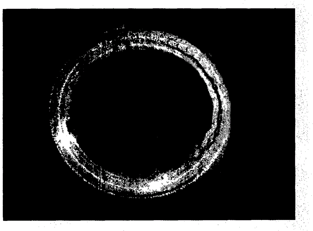
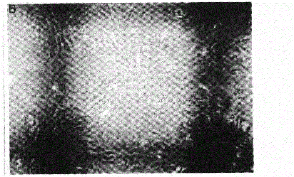
[0044] Severe corneal epithelial defects in rats after alkali burns (see Figure 3a , Figure 3b ), four weeks after the operation, the experimental animal cornea was observed, and each rat was evaluated from five aspects of corneal transparency, corneal turbidity, hyphema, hypopyon, and corneal perforation. The evaluation results are shown in Table 1. It can be seen from Table 1 that the corneal recovery effect after mesenchymal stem cell transplantation is basically the same as that after corneal limbal stem cell transplantation, and is better than simple amniotic membrane transplantation and fibroblast transplantation. In addition, through slit lamp photography, it can be seen that the new blood vessels in the burnt cornea transplanted with mesenchymal stem cells are significantly reduced, the epithelium is basically restored, and the cornea is transparent. The effect is basically the same as that of tran...

Embodiment 2

[0047] Embodiment two, the recovery of burnt rat visual acuity

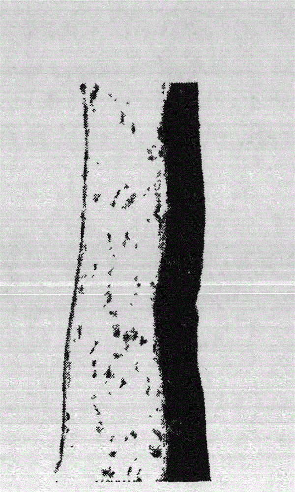
[0048] By optokinetic tracker to the tracking of experimental rat's vision reflex state, the total distance of motion and the trajectory of the single eye before the burn, the single eye before the burn, and the single eye after the burn are compared (see Figure 6a , Figure 6b , Figure 6c , Figure 6d , Figure 6e , Figure 6f ), it can be seen that there is a significant difference in the visual tracker tracking the visual status of rats, and the optokinetic tracker works normally. After the burn, different types of cells were transplanted, and the data of each group were compared horizontally, and the P values ​​were compared through the optokinetic responses of different gratings (see Table 2). There is a significant improvement. MSC: transplanted mesenchymal stem cells; LSC: transplanted limbal stem cells; AM: transplanted amniotic membrane alone; FB: transplanted fibroblasts; CON: negative control. ...

Embodiment 3

[0057] Example 3. Response of inflammatory factors

[0058] MMP-2 (matrix metalloproteinase 2) uses basement membrane collagen as a specific substrate, and its tissue-type inhibitor can directly regulate the enzyme activity in vivo, so MMP-2 can respond to the changes of its new blood vessels in corneal injury. It can be seen from the figure (see Figure 7a , Figure 7b ), the cornea transplanted with mesenchymal stem cells ( Figure 7b ) than the cornea with simple amniotic membrane transplantation ( Figure 7a ) stromal activity is low, indicating that mesenchymal stem cells play a decisive role in inhibiting the regeneration of new blood vessels, showing that the inflammatory response of the corneal stroma of rats transplanted with mesenchymal stem cells is weaker than that of other cells transplanted.


PUM


Abstract

The invention discloses a preparation method of an autologous mesenchymal stem cell-loaded human amniotic membrane cornea paster, wherein autologous sourced mesenchymal stem cells are planted onto an amniotic membrane to be taken as bearing carriers to treat the corneal injury. The method comprises the following steps of: inoculating monocyte separated from bone marrow into an alpha-MEM culture medium, amplifying to a third generation to inoculate 2.5*10<5> cells onto the 1.2cm*1.2cm amniotic membrane, and tightly arranging the cells after 48 hours, wherein the cover area of the mesenchymal stem cell under an inverted microscope is greater than 80%, and the immunofluorescent staining shows that the positive rate of cells CD44 and CD90 is greater than 90%. The human amniotic membrane-loaded mesenchymal stem cell paster is free from dissolving and falling after being transplanted within one week, and new vessels can not be generated within eight months. The method is free from immunological rejection, an enough cell source can be provided, and the method can be used for the clinic treatment of the corneal injury.

Description

technical field [0001] The present invention relates to the field of regenerative medicine, in particular to a preparation method for planting autologous bone marrow mesenchymal stem cells on human amniotic membrane to form a patch for treating corneal damage. Background technique [0002] The treatment of corneal injury is a complicated clinical problem. The cornea is divided into five layers, namely the corneal epithelium, the anterior limiting layer, the corneal stroma, the posterior limiting layer, and the corneal endothelium. Clinical corneal diseases include physical and chemical burns, mechanical trauma, infection, recurrent pterygium, Steven-Johnson syndrome, pekoidoid, trachoma, etc. Severe conjunctival and corneal diseases are still a major part of ophthalmologists’ eyesight treatment. problem. After corneal burns, a large number of limbal stem cells are lost, the corneal epithelium cannot be repaired normally, the cornea is cloudy, new blood vessels increase and...

Claims


Application Information

Patent Timeline
IPC IPC(8): A61L27/38A61L27/36C12N5/0775
Inventor 田杰
Owner 北京清美联创干细胞科技有限公司
Learn More