An imaging method of tumor targeting based on enhancement effect of zinc ion signals
A tumor-targeted imaging and signal enhancement technology, applied in the field of medical imaging, can solve problems such as human hazards, and achieve the effects of low biological toxicity, improved autofluorescence, and easy operation
- Summary
- Abstract
- Description
- Claims
- Application Information
AI Technical Summary
Problems solved by technology
Method used
Image
Examples
Embodiment 1
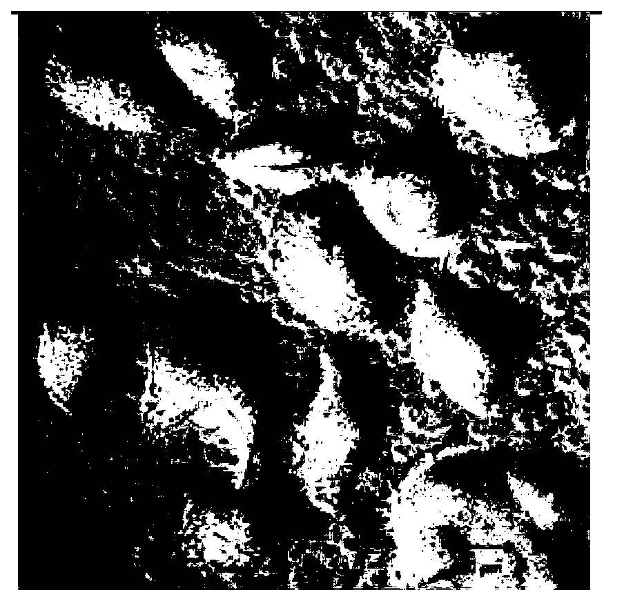
[0028] Dissolve zinc gluconate in PBS solution with pH=7.4 to make 1mmol / L zinc gluconate solution, take hepatoma cell (HepG2) as the research object, put hepatoma cell (HepG2) and zinc gluconate solution in cell culture After incubating at 37°C in the box for 12 hours, the intensity and distribution of cell autofluorescence were detected with a confocal fluorescence microscope, and Raman imaging was performed on liver cancer cells (HepG2) through a Raman microscope, and the signal data of the fluorescence spectrum and the Raman spectrum were used to determine Qualitative and quantitative analysis of the structure or chemical composition of hepatocellular carcinoma cells (HepG2).
Embodiment 2
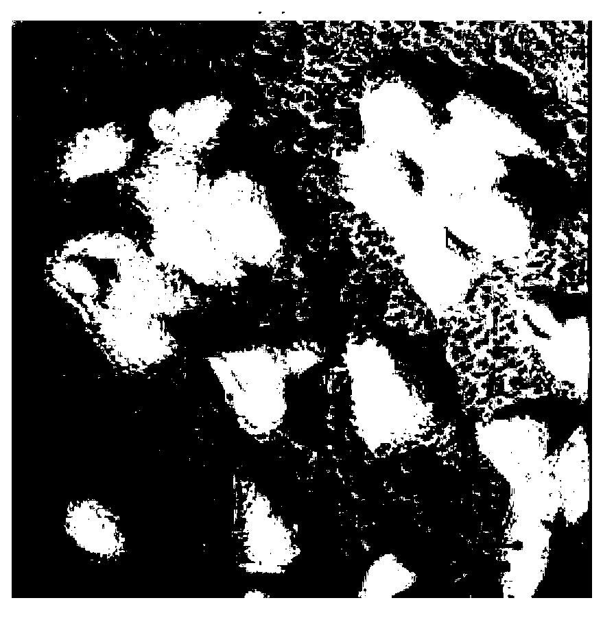
[0030] Dissolve zinc phosphate in PBS solution with pH = 7.4 to make 50mmol / L zinc phosphate solution, take cervical cancer cells (HeLa) as the research object, put cervical cancer cells (HeLa) and zinc phosphate solution in the cell culture incubator After incubating at 37°C for 24 hours, the intensity and distribution of cell autofluorescence were detected with an ultrasonic imager, Raman imaging was performed on cervical cancer cells (HeLa) through a Raman microscope, and the signal data of the fluorescence spectrum and the Raman spectrum were compared. Qualitative and quantitative analysis of cervical cancer cell (HeLa) structure or chemical composition.
Embodiment 3
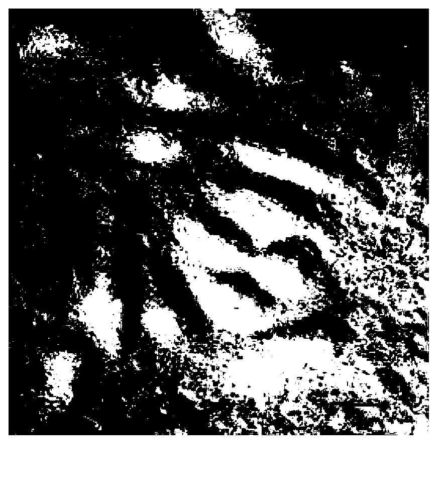
[0032] Dissolve zinc chloride in PBS solution with pH=7.4 to make 100mmol / L zinc chloride solution, take leukemia cells (K562) as the research object, put leukemia cells (K562) and zinc chloride solution in cell culture After incubating at 37°C in the box for 48 hours, use a fluorescence imager to detect the intensity and distribution of cell autofluorescence, use a Raman spectrometer to perform Raman imaging on leukemia cells (K562), and use the signal data of the fluorescence spectrum and Raman spectrum to compare Qualitative and quantitative analysis of the structure or chemical components of leukemia cells (K562).
PUM
 Abstract
Description
Claims
Application Information
 - R&D
- Intellectual Property
- Life Sciences
- Materials
- Tech Scout
- Unparalleled Data Quality
- Higher Quality Content
- 60% Fewer Hallucinations
Browse by: Latest US Patents, China's latest patents, Technical Efficacy Thesaurus, Application Domain, Technology Topic, Popular Technical Reports.
© 2025 PatSnap. All rights reserved.Legal|Privacy policy|Modern Slavery Act Transparency Statement|Sitemap|About US| Contact US: help@patsnap.com


