Electrochemiluminescent imaging device and applications thereof

A technology of electrochemistry and luminescence imaging, which is applied in the direction of chemiluminescence/bioluminescence, measuring devices, and analysis by making materials undergo chemical reactions, which can solve the problems of detection flux limitation and impossibility, and achieve simple composition , increase the flux, promote the effect of luminous efficiency

Active Publication Date: 2014-07-09
NANJING MEDICAL UNIV
View PDF8 Cites 7 Cited by
  • Summary
  • Abstract
  • Description
  • Claims
  • Application Information

AI Technical Summary

Problems solved by technology

The disadvantage of this technology is that each cell surface must be detected independently, so the detection throughput is severely limited
Although in the presence of luminol, these released molecules are converted into electrochemiluminescent hydrogen peroxide by its oxidase, but it must be realized in the submicron region, and the sampling time of the second level (time In the range of resolution), it is still impossible to obtain a detectable optical signal for such a low concentration of biomolecules

Method used


Image

  • Electrochemiluminescent imaging device and applications thereof
  • Electrochemiluminescent imaging device and applications thereof
  • Electrochemiluminescent imaging device and applications thereof

Examples


Embodiment 1

[0035] The visual detection of hydrogen peroxide in the solution of embodiment 1.

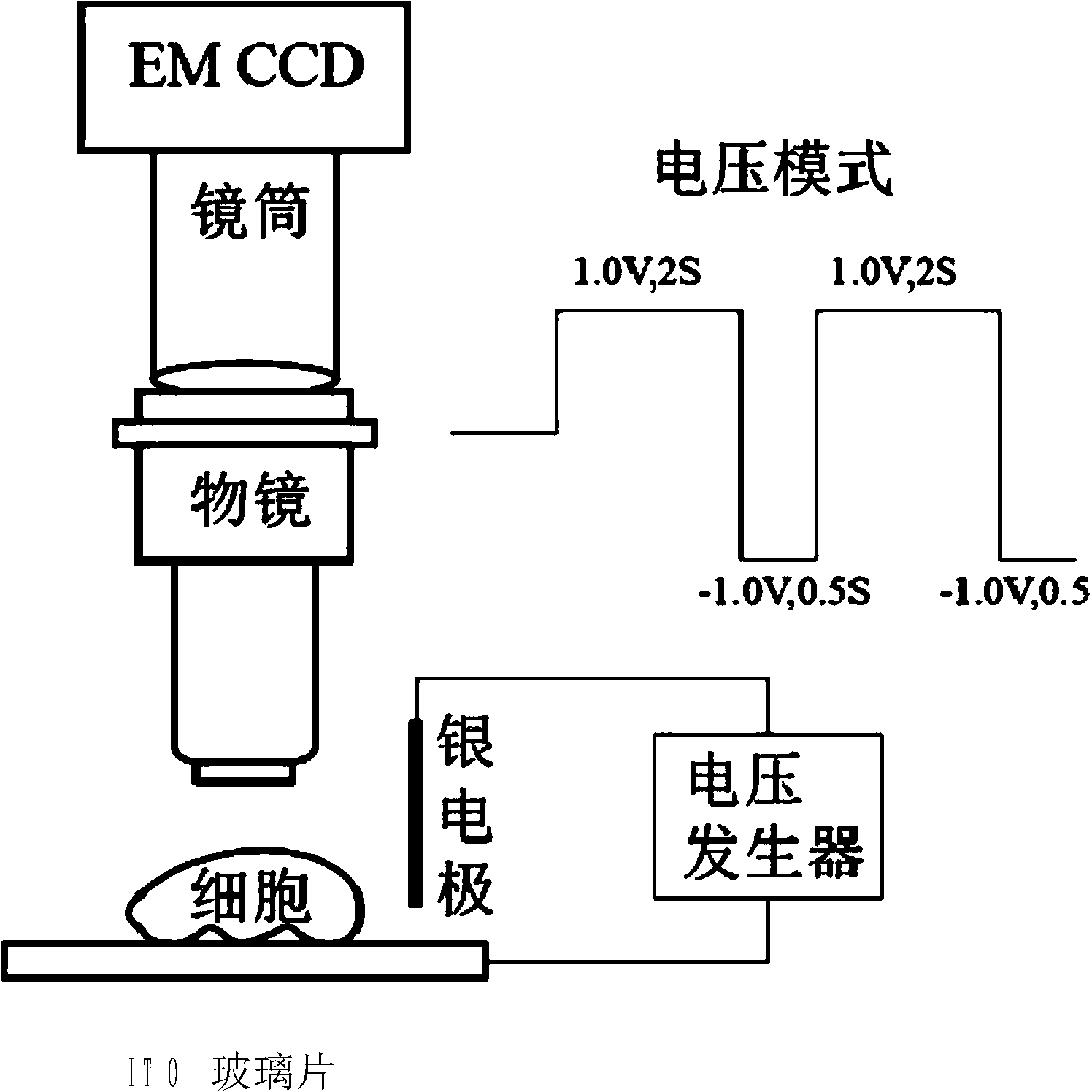
[0036] (1) Determination of detection conditions.

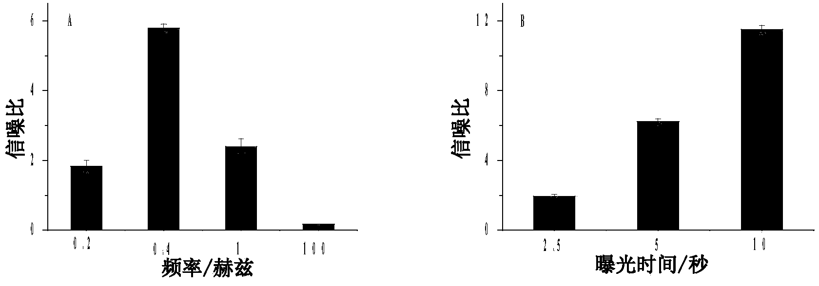
[0037] In the experiment, an O-ring with a diameter of 1 cm was pasted on an indium tin oxide (ITO) glass sheet as a solution chamber, and the ITO glass sheet was used as a working electrode, and L012 was selected as the luminescent substance under positive voltage. L012 is a luminol analog, which has a higher luminescence effect than luminol in the presence of hydrogen peroxide, so it was selected as the luminescence substance under positive voltage. Add 200 μM L012 and 100 μM hydrogen peroxide dropwise into the O-ring, totaling 600 μL. In view of the luminescence peak voltage of L012 and hydrogen peroxide on the ITO glass electrode is 1.0V (wherein, 1.0V is the peak voltage of electrolysis of hydrogen peroxide on the ITO electrode surface, and then the oxygen free radicals produced make L012 luminescence, so for different luminescent reagen...

Embodiment 2

[0043] Example 2 Visual detection of hydrogen peroxide leakage from the surface of single cells.

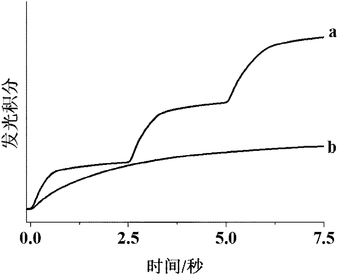
[0044] Single-cell leaked hydrogen peroxide imaging was performed by culturing 100-5000 Hela cells in a solution chamber on an ITO glass slide, and then using 10 μl of 20ng / ml crotyl alcohol-12-myristate-13-acetate (PMA, Phorbol12 -myristate-13-acetate, referred to as phorbol ester, purchased from sigma-aldrich, product number is P8139) stimulates the production of hydrogen peroxide. According to reports, PMA stimulates NADPA oxidase in cells to accumulate hydrogen peroxide, which leads to the leakage of hydrogen peroxide. According to the same imaging steps, the bright field image and the background image ( Figure 5 A and 5B). In the luminescence map, due to the fact that the cells attached to the electrode surface hindered the diffusion of L012 to the electrode surface, it appeared as a darker intensity. Immediately after the cells released hydrogen peroxide after PMA stimu...

Embodiment 3

[0047] Example 3 Electrochemiluminescence imaging of molecules on the surface of single cells.

[0048]In addition to imaging the hydrogen peroxide released by cells, the electrochemiluminescence imaging method of the present invention can also convert molecules on the cell surface into hydrogen peroxide by oxidase corresponding to them, thereby realizing the imaging of the molecules on the cell surface. In order to demonstrate the possibility of imaging molecules on the cell surface, we chose cholesterol as a template molecule to detect the distribution of activated cholesterol molecules on cell membranes. Activated cholesterol has a high chemical potential (escape tendency), which plays an important role in cell membrane cholesterol transport. Although cell membrane cholesterol has been imaged using fluorescent cholesterol analogues or flipins, activated cholesterol on the cell surface has never been successfully imaged because these fluorescent probes cannot distinguish act...


PUM


Abstract

The invention discloses an electrochemiluminescent imaging device which comprises an object lens, a light transmission channel, a single-photon detection electron multiplication CCD (charge coupled device) and an electrochemiluminescent generator, wherein the electrochemiluminescent generator comprises an ITO (indium tin oxide) electrode, a silver electrode and a voltage generator; the object lens, the light transmission channel and the single-photon detection electron multiplication CCD are connected in sequence; the ITO electrode is connected with the positive electrode of the voltage generator; the silver electrode is connected with the negative electrode of the voltage generator. The invention further discloses applications of the electrochemiluminescent imaging device to visual detection on hydrogen peroxide, hydrogen peroxide leaking from the surface of single cells and cell surface molecules. The electrochemiluminescent imaging device is simple in composition, high in detection flux, easy to operate and high in luminous efficiency, and can perform the parallel quick detection.

Description

technical field [0001] The invention belongs to the technical field of biochemical industry, and in particular relates to an electrochemiluminescence imaging device and its application. Background technique [0002] Imaging studies on single cells using electrochemical techniques can obtain important biological information such as chemical components and local activities on the cell surface. Currently, the most common microscopic instrument is an electrochemical scanning microscope. The microscope uses a microelectrode to scan the surface of a single living cell to detect electrically active chemical species. By reducing the electrode size to the nanometer level, the spatial resolution of the electrochemical scanning microscope can reach the nanometer level. The disadvantage of this technology is that each cell surface must be detected independently, so the detection throughput is severely limited. As an alternative to electrochemical scanning microscopy, the recently deve...

Claims


Application Information

Patent Timeline
Patent Type & Authority Applications(China)
IPC IPC(8): G01N21/76G01N21/69G01N21/01
Inventor 方丹君江德臣周俊宇
Owner NANJING MEDICAL UNIV
Why Eureka
Try Eureka