An electrochemiluminescence imaging device and its application
A technology of electrochemistry and luminescence imaging, which is applied in the direction of chemiluminescence/bioluminescence, measuring devices, and analysis by making materials undergo chemical reactions, which can solve the problems of detection flux limitation and impossibility, and achieve simple composition , increase the flux, promote the effect of luminous efficiency
- Summary
- Abstract
- Description
- Claims
- Application Information
AI Technical Summary
Problems solved by technology
Method used
Image
Examples
Embodiment 1
[0035] Example 1 Visual detection of hydrogen peroxide in solution.
[0036] (1) Determination of detection conditions.
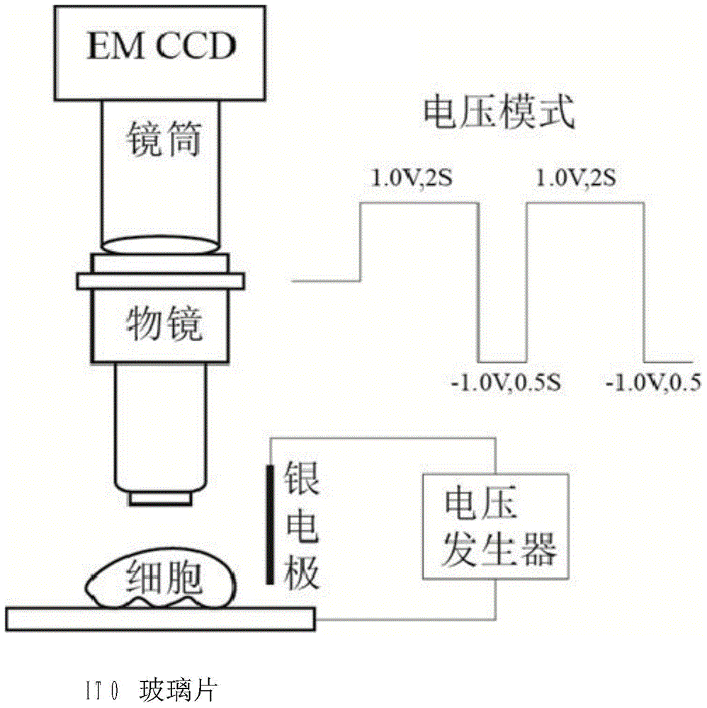
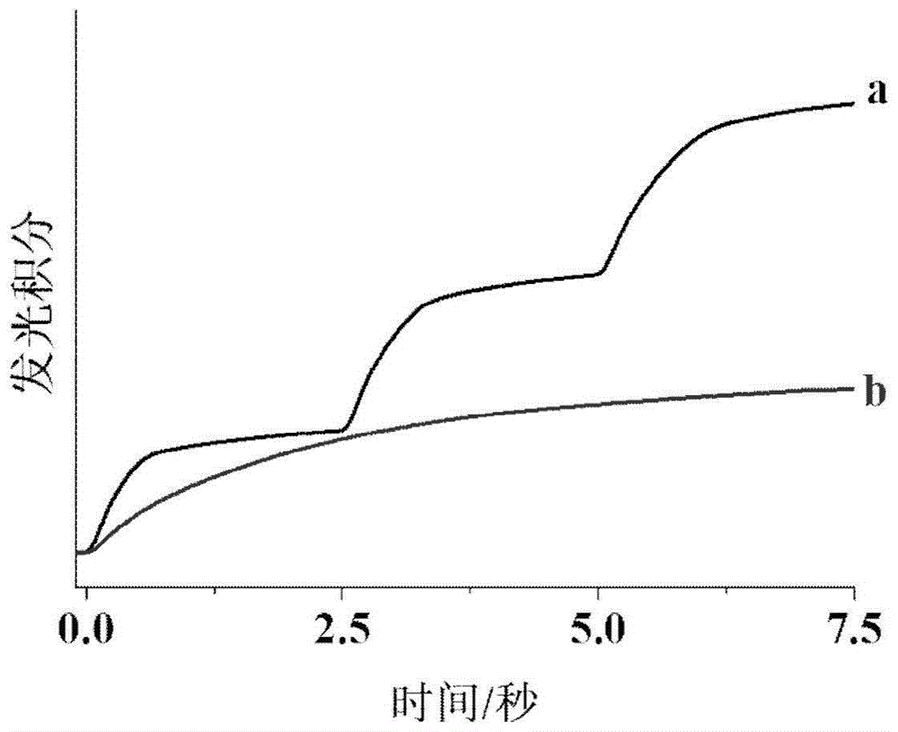
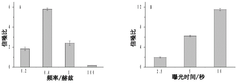
[0037] In the experiment, an O-ring with a diameter of 1 cm was pasted on an indium tin oxide (ITO) glass sheet as the solution chamber, and the ITO glass sheet was used as the working electrode, and L012 was selected as the luminescent substance under positive voltage. L012 is a luminol analog, which has a higher luminescence effect than luminol in the presence of hydrogen peroxide, so it was selected as the luminescence substance under positive voltage. 200 μM of L012 and 100 μM of hydrogen peroxide were added dropwise to the O-ring for a total of 600 μL. In view of the fact that the luminescence peak voltage of L012 and hydrogen peroxide on the ITO glass electrode is 1.0V (wherein, 1.0V is the peak voltage of the electrolysis of hydrogen peroxide on the surface of the ITO electrode, and then the generated oxygen radicals make L012 emit light, so for dif...
Embodiment 2
[0043] Example 2 Visual detection of hydrogen peroxide leakage from the surface of single cells.
[0044] Single-cell leakage of hydrogen peroxide imaging was performed by culturing 100-5000 HeLa cells in solution chambers on ITO glass slides and then treated with 10 μl of 20 ng / ml crotyl alcohol-12-tetradecanoate-13-acetate (PMA, Phorbol12 -myristate-13-acetate, referred to as phorbol ester, purchased from sigma-aldrich, item number is P8139) to stimulate the production of hydrogen peroxide. It has been reported that PMA stimulates intracellular NADPA oxidase to accumulate hydrogen peroxide, resulting in the leakage of hydrogen peroxide. According to the same imaging procedure, brightfield and background images in the presence of L012 were obtained ( Figure 5 A and 5B). In the luminescence image, it appears as a darker intensity due to the adhesion of cells to the electrode surface, which hinders the diffusion of L012 to the electrode surface. After the cells released hyd...
Embodiment 3
[0047] Example 3 Electrochemiluminescence imaging of single cell surface molecules.
[0048]In addition to imaging the hydrogen peroxide released by the cells, the electrochemiluminescence imaging method of the present invention can also utilize the oxidase corresponding to the molecules on the cell surface to convert them into hydrogen peroxide, thereby realizing the imaging of the cell surface molecules. To demonstrate that cell surface molecules can be imaged, we chose cholesterol as a template molecule to detect the distribution of activated cholesterol molecules in the cell membrane. Activated cholesterol has a higher chemical potential (tendency to escape), which is important for cholesterol transport across cell membranes. Although cell membrane cholesterol has been imaged using fluorescent cholesterol analogs or flipins, cell-surface activated cholesterol has never been successfully imaged because these fluorescent probes cannot differentiate between activated and non-...
PUM
 Abstract
Description
Claims
Application Information
 - R&D
- Intellectual Property
- Life Sciences
- Materials
- Tech Scout
- Unparalleled Data Quality
- Higher Quality Content
- 60% Fewer Hallucinations
Browse by: Latest US Patents, China's latest patents, Technical Efficacy Thesaurus, Application Domain, Technology Topic, Popular Technical Reports.
© 2025 PatSnap. All rights reserved.Legal|Privacy policy|Modern Slavery Act Transparency Statement|Sitemap|About US| Contact US: help@patsnap.com


