Solid fluorescent nanometer microsphere as well as preparation method and application thereof

A fluorescent nanometer and solid technology, applied in the field of fluorescence detection, can solve the problems of weak fluorescence and poor uniformity of surface fluorescent microspheres, and achieve the effects of uniform fluorescence intensity, high signal amplification, and improved sensitivity.

Active Publication Date: 2014-07-23
WUHAN NEWCANDO BIOTECH CO LTD
View PDF3 Cites 15 Cited by
  • Summary
  • Abstract
  • Description
  • Claims
  • Application Information

AI Technical Summary

Problems solved by technology

[0005] The usual fluorescent microspheres are mostly surface fluorescent microspheres, which are made by coupling fluorescent molecules to the surface of the microspheres through specific functional groups. The surface fluorescent microspheres have weak fluorescence and poor uniformity.

Method used


Image

  • Solid fluorescent nanometer microsphere as well as preparation method and application thereof
  • Solid fluorescent nanometer microsphere as well as preparation method and application thereof
  • Solid fluorescent nanometer microsphere as well as preparation method and application thereof

Examples


Embodiment 1

[0039] Embodiment 1: Synthesis of Entity Fluorescent Nanospheres

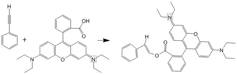
[0040] 1. Dissolve 1-10 mmol of Rhodamine B in 10 mL of DMSO.

[0041] 2. Dissolve 1-10mmol of phenylacetylene in 10mL of DMSO, and then mix it with Rhodamine B in DMSO.

[0042] 3. Add 20mL of 1-10mmol CuCl to the reaction system in step 2 2 Aqueous solution, adding 50-300mL of water to the mixed solution, stirring and reacting at room temperature for 2-4 hours at a speed of 300rpm under nitrogen protection.

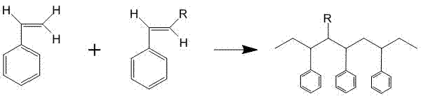
[0043] 4. Add 0.5-5g sodium dodecylsulfonate (SDS) as an emulsifier, 0.29-2.9g hydroxydicumyl peroxide (CHPO) and 0.23-2.3g tetraethylenepentamine to the reaction system in step 3 (TEPA) as oxidation-reduction initiator, 10mL DMSO solution of 5~50mmol styrene as reactant, pass into nitrogen (N 2 ), continuously stirred at a speed of 1000-5000rmp, and polymerized at 42°C for 2-4 hours.

[0044] 5. Centrifuge the obtained polymer at a rotational speed of 8000-10000 rpm, take the precipitate, wash with ...

Embodiment 2

[0051] Example 2 Preparation of Entity Fluorescent Nanospheres

[0052] 1. Dissolve 0.1g Rhodamine B in 200μL DMSO.

[0053] 2. Dissolve 0.1 g of phenylacetylene in 200 μL of DMSO, and then mix with Rhodamine B in DMSO.

[0054] 3. Add 5 mL of water to the reaction system, and stir for 2 hours at room temperature.

[0055] 4. Add 0.5g sodium dodecylsulfonate (SDS) to the reaction system as an emulsifier, 0.29g hydroxydicumyl peroxide (CHPO) and 0.23g tetraethylenepentamine (TEPA) as an oxidation-reduction initiator Agent, 0.5g styrene as reactant, pass N 2 , continuously stirred at 5000rmp, and polymerized at 42°C for 2h.

[0056] 5. The obtained polymer is washed with ethanol and then vacuum-dried to obtain the product.

Embodiment 3

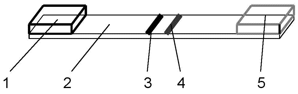
[0057] Example 3: Making an immunochromatographic detection reagent for solid fluorescent nanosphere NT-proBNP

[0058] 1. Labeled antibody:

[0059] (1) Take 0.5mL solid fluorescent nanospheres, centrifuge to get the precipitate, add MES buffer with pH 6.0 to wash twice.

[0060] (2) Activation: Add 200 μL of 50 mM carbodiimide (EDAC) and 200 μL of 50 mM hydroxysulfosuccinimide (Sulfo-NHS) to the washed microspheres, shake at room temperature for 2 h.

[0061] (3) Centrifuge the microspheres and wash twice with PBS.

[0062] (4) Add an appropriate amount of antibody and shake at room temperature for 2 hours.

[0063] (5) Centrifuge the microspheres, add 50mM glycine to quench the reaction, and shake at room temperature for 5 minutes; then centrifuge the microspheres, add 50mM glycine to completely quench the reaction, and shake at room temperature for 15 minutes.

[0064] ⑹ Centrifuge the microspheres, add blocking solution (1% BSA, 5% sucrose, 0.2% Tween-20, 0.1% sodium a...


PUM


Abstract

The invention discloses a solid fluorescent nanometer microsphere as well as a preparation method and an application thereof, and belongs to the technical field of fluorescence detection. The solid fluorescent nanometer microsphere disclosed by the invention is prepared from rhodamine B-styrene which is prepared by carrying out covalent addition on carboxyl rhodamine B and phenylacetylene and polymerizing the rhodamine B-styrene and styrene according to the molar ratio of 1:(1-12). A fluorescent molecule is embedded into a space network of polystyrene, and the obtained solid fluorescent microsphere has the characteristics of being uniform and consistent in fluorescence intensity among the microspheres, high in fluorescence intensity, high in signal amplification factor and the like. No hinder of the fluorescent molecule exists on the surface of the microsphere, so that the microsphere can be coupled to protein with the high efficiency and the high productivity. The fluorescent background generated by the microsphere is low, the accuracy of the detection result is not affected, and the solid fluorescent nanometer microsphere can be widely applied to a diagnostic reagent.

Description

technical field [0001] The invention relates to the technical field of fluorescence detection, in particular to a solid fluorescent nano-microsphere, a preparation method thereof and an application in immunoquantitative chromatography. Background technique [0002] Lateral flow chromatography has been widely used in the fields of rapid diagnosis of diseases and rapid detection of small molecules. The principle is to coat the specific antigen or antibody on the nitrocellulose membrane to form a detection line, and to coat the secondary antibody in an area 2 to 3 mm away from the detection line to form a quality control line. After the target molecules in the sample are combined with the detection antibody with a specific label, the nitrocellulose membrane is chromatographed from the sample-loading side to the water-absorbing side. Due to capillary action, the complex will move forward along the membrane, but when moving to the detection line, the target molecules in the samp...

Claims


Application Information

Patent Timeline
Patent Type & Authority Applications(China)
IPC IPC(8): G01N21/64G01N33/558B82Y40/00
Inventor 郑忠亮吴琼水蔡磊朱辉
Owner WUHAN NEWCANDO BIOTECH CO LTD
Why Eureka
Try Eureka