Application of indocyanine green-rituximab in preparation of breast cancer sentinel node tracer

A technology of rituximab and sentinel lymph node, which is applied in the field of specific tracers, can solve the problems of complex preparation process, radionuclide pollution, and inaccurate imaging, and achieve high sensitivity, difficulty in dissociation, and clear imaging. The effect of high image rate

Active Publication Date: 2015-05-20
王永胜
View PDF2 Cites 3 Cited by
  • Summary
  • Abstract
  • Description
  • Claims
  • Application Information

AI Technical Summary

Problems solved by technology

Among them, the dye method (methylene blue, patent blue and nano-carbon) locates the sentinel lymph node mainly through the preoperative dye around the primary breast tumor and (or) subcutaneously injecting the dye, and then following the dye-stained lymphatic vessels in the axillary area for fine dissection Find the first stained lymph node to determine the specific location of the sentinel lymph node. However, this method requires superb surgical anatomy skills, and there is still a high false-negative rate of sentinel lymph node biopsy after the learning curve, which leads to sentinel lymph node biopsy. The accuracy of the method is difficult to grasp; while the nuclide method ( 99m Tc-sulfur colloid) is used to locate the sentinel lymph node by preoperatively injecting radionuclide into the glands around the primary breast tumor and (or) subcutaneously, and then use the gamma detector to detect the radioactive count during the operation, and find the lymph node with a high count according to the radioactive count , so as to determine the specific location of the sentinel lymph node (lymph node greater than 10% of the highest radioactive count), although this method of sentinel lymph node biopsy has high accuracy and good specificity, but there are risks of radionuclide contamination and radiation damage to medical staff during actual operation problems, and the sulfur colloid has not been approved by the China Food and Drug Administration, which limits its promotion and use in clinical practice; the method of locating sentinel lymph nodes by fluorescence method (indocyanine green) is similar to the dye method, mainly After the preoperative injection of a fluorescent tracer around the primary breast tumor and / or subcutaneously, the first lymph node that is positive for fluorescence imaging is found by using a near-infrared fluorescence imaging device to follow the bright lymph vessels of fluorescence imaging during surgery , so as to locate the specific location of the sentinel lymph node. However, since the fluorescent tracer molecules need to be excited by a near-infrared light source, the penetration of the fluorescent light released by the excited is only about 9mm, so it is difficult to find the deep lymphatic vessels during the biopsy , resulting in the missed detection of sentinel lymph nodes. Although the shallower lymphatic vessels can be clearly detected by fluorescence imaging, the fluorescent tracer in them leaks out together with the lymph fluid after they are disconnected, which can easily cause fluorescence in the tissues around the lymphatic vessels. This is not an ideal sentinel lymph node tracer.
In addition, current specific sentinel lymph node imaging agents 99m The preparation process of Tc-rituximab is relatively complicated, and it needs to go through steps such as antibody modification, separation after modification, antibody packaging, antibody labeling, separation and purification, etc. Exposure to radioactive substances can easily cause radiation damage to experimental technicians, surgeons, pathologists and other related personnel. In addition, the labeling rate of rituximab and radionuclides prepared by this method is sometimes only 90%, which is difficult to meet Sentinel lymph node imaging and positioning requirements may easily lead to inaccurate imaging
[0003] The currently existing combination products of rituximab and indocyanine green are only used for the diagnosis of ex vivo cell detection, and have not been applied in the imaging and localization of sentinel lymph node biopsy of breast cancer, and the combination of rituximab and indocyanine green The preparation method of docyanine green combination has no detailed operation steps and specific dosage instructions, and there are currently no reports and studies on the application of sentinel lymph node biopsy

Method used


Image

  • Application of indocyanine green-rituximab in preparation of breast cancer sentinel node tracer
  • Application of indocyanine green-rituximab in preparation of breast cancer sentinel node tracer
  • Application of indocyanine green-rituximab in preparation of breast cancer sentinel node tracer

Examples


Embodiment 1

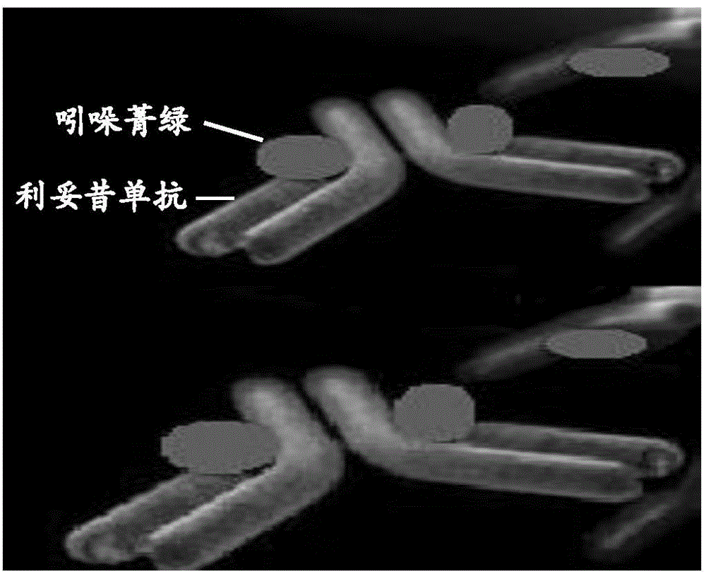
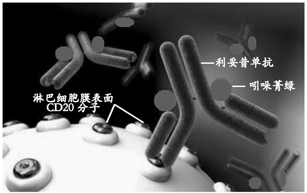
[0035] Application of indocyanine green-rituximab in preparation of sentinel lymph node tracer for breast cancer.

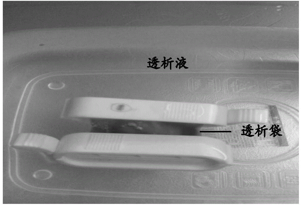
[0036] At room temperature, according to the principle of aseptic operation, 100mg of rituximab (Rituxan 100mg Roche) was made into 10mg / ml with 10ml of sterile water for injection (5ml of sterile water for injection, Tianjin Pharmaceutical Group Xinzheng Co., Ltd.) The solution is placed in a 50ml sterile centrifuge tube and stored under refrigeration (2-8°C) for later use; 25mg indocyanine green (indocyanine green for injection 25mg Dandong Medical Chuangye) is formulated with 10ml sterile water for injection to a concentration of The 2.5mg / ml solution should be stored in a 50ml sterile centrifuge tube and protected from light for later use. At room temperature, take 6 portions of 0.5ml of the prepared 10mg / ml rituximab solution and place them in a 10ml centrifuge tube, and then take the mass ratio of rituximab to indocyanine green to 3 : 1, 4:1, 6:1, 12:1, 30:1 ...

Embodiment 2

[0037] Example 2 Detecting the molecular integrity of the indocyanine green-rituximab coupling reactant:

[0038] 8% non-reducing SDS polyacrylamide gel electrophoresis was used to detect the molecular integrity of indocyanine green-rituximab, and rituximab was used as a positive control to determine indocyanine green-rituximab Molecular integrity, using the coupling reactants with mass ratios of rituximab and indocyanine green of 4:1, 6:1, 12:1, 30:1, and 32:1 in Example 1 did not appear Obviously degraded bands, such as Figure 4 As shown (the gel spectra of the coupling reactants in the above mass ratios are consistent, and no degradation bands are seen), it shows that the molecular structure of the coupling products of the two is complete.

Embodiment 3

[0039] Example 3 Detection of the immunological activity of the indocyanine green-rituximab conjugate:

[0040] Double antibody sandwich indirect enzyme-linked immunoassay (ELISA) method to detect the immune activity of indocyanine green-rituximab, rabbit anti-mouse IgG-Fab antibody is a coating antibody, and the mass fraction is 1% bovine serum albumin blocked For 1 hour, add the conjugates with mass ratios of rituximab to indocyanine green of 4:1, 6:1, 12:1, 30:1 and 32:1 in Example 1, and avoid at room temperature. Light reaction for 2 hours, add horseradish peroxidase-labeled goat anti-human IgG-Fc antibody, and react for 1 hour in the dark at room temperature. The color is developed by o-phenylenediamine hydrochloride. The enzyme-linked analyzer measures A 412nm Value such as Figure 5 As shown, the results show that the color reaction of the coupling reactants with mass ratios of rituximab and indocyanine green of 4:1, 6:1, 12:1, 30:1 and 32:1 was positive, and the OD 412 ...


PUM


Abstract

The invention discloses an application of indocyanine green-rituximab in preparation of a breast cancer sentinel node tracer. When sentinel node biopsy is carried out via an indocyanine green-rituximab coupled specific tracer, since the specific tracer only fluorescently develops the sentinel node and develops no secondary lymph node, the sentinel node does not need to be found along the fluorescently developed lymphatic in the biopsy, the position of the fluorescently developed sentinel node is detected by an infrared fluorescent development system in a surgery, and the sentinel node positioned by fluorescent development is directly dissected and detected after dissecting and dissociating the peripheral tissues of the sentinel node, so as to avoid the situations of leak detection of the sentinel node and fluorescent development pollution of the peripheral tissues.

Description

Technical field [0001] The invention relates to an application of indocyanine green-rituximab to prepare a specific tracer for breast cancer sentinel lymph node. Background technique [0002] The sentinel lymph node is the first lymph node of breast cancer metastasis, and its pathological condition has important guiding value for the surgical operation and comprehensive treatment of breast cancer. Sentinel lymph node biopsy is a technical method for breast cancer lymph node staging, and accurate positioning of the sentinel lymph node is a technical field that has been focused on in the research of breast cancer sentinel lymph node biopsy. Traditional tracers for sentinel lymph node location include dyes, nuclides, fluorescence, etc., which are all non-specific tracers. Among them, the dye method (methylene blue, patent blue and nano carbon) locates the sentinel lymph node mainly by injecting the dye around the primary breast tumor and / or subcutaneously before the operation, and ...

Claims


Application Information

Patent Timeline
Patent Type & Authority Applications(China)
IPC IPC(8): A61K49/00
Inventor 王永胜丛斌斌孙晓宋现让杨国仁曹晓珊田崇麟张燕李盼盼
Owner 王永胜
Why Eureka
Try Eureka