3D (three-dimensional) printing type porous metal scaffold with superficial nanocomposite coatings and preparation of 3D printing type porous metal scaffold

A nano-composite coating, porous stent technology, applied in coatings, electrolytic coatings, prostheses, etc., can solve the problems of increased contact area, low biological activity on the alloy surface, implantation failure, etc., and achieves low cost, film layer Good morphology uniformity and improved biological activity

Inactive Publication Date: 2015-12-09
PEKING UNIV
View PDF2 Cites 20 Cited by
  • Summary
  • Abstract
  • Description
  • Claims
  • Application Information

AI Technical Summary

Problems solved by technology

Compared with bulk matrix materials, the surface area of ​​EBM3D porous Ti6Al4V scaffold and the contact area with bone cells are significantly increased, and it is more vulnerable to the erosion of the liquid environment in the human body, resulting in the dissolution of Al and V elements, resulting in cytotoxicity and immune reactions. At the same time, Ti6Al4V The surface of the alloy has low bioactivity and cannot form a firm bone bond with bone tissue, resulting in implant failure

Method used


Image

  • 3D (three-dimensional) printing type porous metal scaffold with superficial nanocomposite coatings and preparation of 3D printing type porous metal scaffold
  • 3D (three-dimensional) printing type porous metal scaffold with superficial nanocomposite coatings and preparation of 3D printing type porous metal scaffold
  • 3D (three-dimensional) printing type porous metal scaffold with superficial nanocomposite coatings and preparation of 3D printing type porous metal scaffold

Examples


Embodiment 1

[0035] Cut silkworm cocoon (Bombyxmoricocoon) into pieces, weigh 5g, and place in 2L0.02MNa 2 CO 3 Boil in the solution for 30 minutes to remove sericin on the surface of silk. After boiling, wash the degummed silk fibroin several times with deionized water and dry it naturally. Then dissolve the silk fibroin in 9MLiBr at 60°C until the silk fibroin is completely dissolved Afterwards (about 4h), dialyze with a dialysis membrane for 3 days to obtain a silk fibroin aqueous solution with a mass volume fraction of 6-9w / v% (g / mL), which is stored in a refrigerator at 4°C until use.

Embodiment 2

[0037] Put the 8w / v% silk fibroin (silk) solution freshly prepared in Example 1 in a 50ml centrifuge tube, tighten the cap and place it in a 60°C water bath for overnight treatment. Prepare the deposition solution according to the composition ratio in Table 1. Among them, in order to ensure that graphene oxide (GO) is fully dispersed in the silk fibroin solution, 200 μL GO solution can be mixed with 4.9 mL deionized water first, and then added to the silk fibroin solution. Hydroxyapatite (HA) nanoparticles (about 40nm in diameter) need to be dispersed in 4.9mL deionized water and 1.1mL ethanol solution, after ultrasonic dispersion for 30min, add HA / deionized water / ethanol solution to silk fibroin solution , stir well. After the deposition solution is prepared, it can be deposited after standing for 20-30 minutes.

[0038] Table 1 The composition of silk fibroin / GO / HA deposition solution

[0039]

Embodiment 3

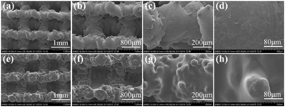
[0041] The 3D printed porous Ti6Al4V scaffolds were ultrasonically cleaned in acetone, 75% ethanol and deionized water for 15 min each, and dried at 60 °C for 1 h. Activate with acid solution at room temperature for 3 minutes. The formula of acid solution is nitric acid: hydrofluoric acid: water = 28.8: 2.4: 70 (volume ratio). The deposition solution in Example 3 was used as the deposition solution, 316L stainless steel as the cathode, and the porous Ti6Al4V support as the anode, the cathode and the anode were placed at a distance of 10mm, a DC voltage of 10V was applied between the two stages, and the deposition time was 30-45s. After the deposition, the samples were soaked in 90% methanol solution for 2 h, and then dried naturally. The surface morphology of the bare stent and the electrophoretic deposition pure silk fibroin coating surface SEM morphology are as follows: figure 1 shown.

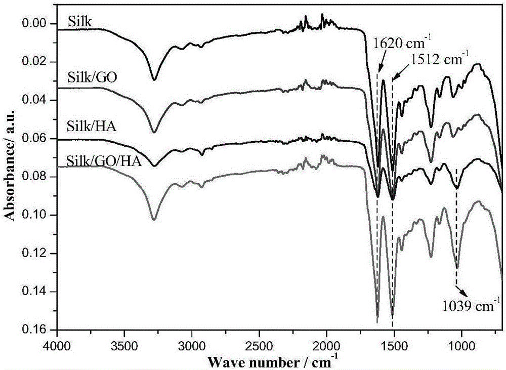
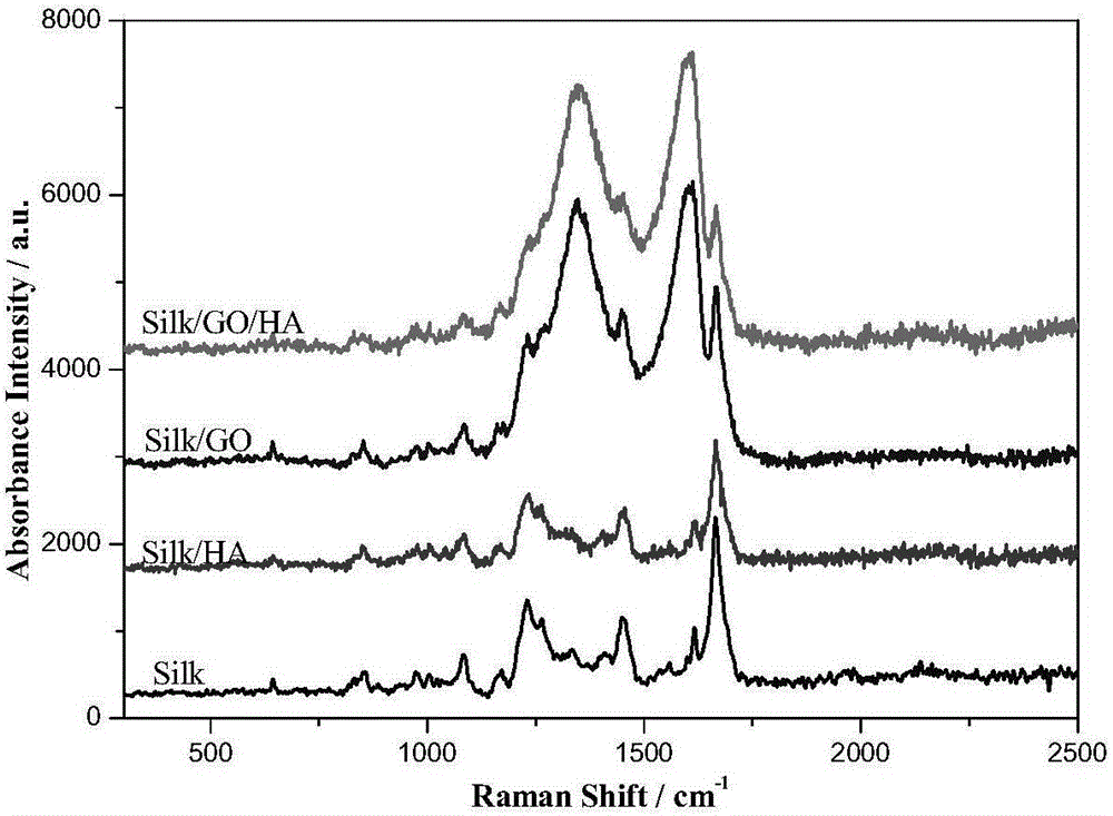
[0042] The materials obtained in Examples 1 to 3 are characterized by scanning electro...


PUM

PropertyMeasurementUnit
Particle sizeaaaaaaaaaa
Mass volume fractionaaaaaaaaaa

Abstract

The invention discloses a 3D (three-dimensional) printing type porous metal scaffold with superficial nanocomposite coatings and a preparation method of the 3D printing type porous metal scaffold and relates to the preparation field of functionalized porous scaffolds for bone tissue repair and reconstruction. The silk fibroin-graphene oxide nanocomposite coatings are prepared on nonlinear metal surfaces of a 3D printing type porous Ti6A14V alloy scaffold and the like by an electrophoretic deposition method or a Czochralski method, and the functional coatings uniform in structure and good in binding force are obtained on the inner and outer surfaces of the scaffold at the same time, so that matrix biological activity is improved and matrix corrosion rate is decreased.

Description

technical field [0001] The invention belongs to the technical field of bone tissue repair and reconstruction, and in particular relates to a preparation method and a functionalized support for a 3D printed porous titanium-based alloy surface functionalized biological coating for bone tissue repair and reconstruction. Background technique [0002] Due to the diversification and complexity of orthopedic diseases, traditional orthopedic implants cannot match the individual differences of patients' bones, and it takes a long operation time to shape and match traditional implants and bone tissue defects, which increases the bleeding of patients during operation Therefore, on the basis of modern imaging and computer-aided design, the use of 3D printing technology to prepare individualized implants for patients can significantly improve the accuracy and safety of orthopedic surgery and meet the needs of personalized treatment for patients. demand. [0003] 3D printing belongs to r...

Claims


Application Information

Patent Timeline
IPC IPC(8): A61L27/34A61L27/06A61L27/54A61L31/10A61L31/02A61L31/16B22F3/105B22F3/115C25D13/12
Inventor 李明成艳郑玉峰
Owner PEKING UNIV
Why Eureka
Try Eureka