Two-photon deep red emission fluorescent probe for imaging cell membranes in tissues based on molecular rotors

A fluorescent probe and two-photon technology, applied in the direction of fluorescence/phosphorescence, luminescent materials, analytical materials, etc., can solve the problems of increasing the difficulty of synthesis and limiting applications, and achieve large Stokes shift, good compatibility, The effect of filling the gap

Active Publication Date: 2016-05-11
SHANDONG UNIV
View PDF5 Cites 10 Cited by
  • Summary
  • Abstract
  • Description
  • Claims
  • Application Information

AI Technical Summary

Problems solved by technology

Undoubtedly, this increases the difficulty of synthesis on the one hand, and on the other hand, such a large molecular structure limits its application in tissue samples

Method used


Image

  • Two-photon deep red emission fluorescent probe for imaging cell membranes in tissues based on molecular rotors
  • Two-photon deep red emission fluorescent probe for imaging cell membranes in tissues based on molecular rotors
  • Two-photon deep red emission fluorescent probe for imaging cell membranes in tissues based on molecular rotors

Examples


Embodiment 1

[0027] Synthesis of N,N-Diformylaniline

[0028] Dry DMF (3.7mL) and phosphorus oxychloride (3.7mL, 40.7mmol) were mixed into a three-necked flask and stirred at 0°C. After 30 min, triphenylamine (1.0 g, 4.0 mmol) dissolved in chloroform was added dropwise to the above mixture, heated and stirred for 24 h. Cool to room temperature after the reaction, add appropriate amount of sodium hydroxide and water, then extract with dichloromethane and wash with water. Dry with anhydrous sodium sulfate. Finally, a mixture of petroleum ether and ethyl acetate was used for column chromatography to obtain the final product.

[0029] 1 HNMR (400MHz, DMSO-d6): δ (ppm) 9.88 (s, 2H), 7.85 (d, J = 8.64Hz, 4H), 7.49 (t, J = 7.84Hz, 2H), 7.33 (t, J = 7.4Hz, 1H), 7.22(d, J=8.4Hz, 2H), 7.17(d, J=8.56Hz, 4H).

[0030] Synthesis of 1-dodecyl-4-picoline-1 iodide

[0031] Mix 4-methylpyridine (2 mL, 20 mmol) and 1-iodododecane, use ethanol as solvent, and heat to reflux. After 24 hours, the reaction...

Embodiment 2

[0036] Example 2: Immortalized cancer cell (HeLa and SiHa) and normal cell (HUVEC) culture

[0037] HeLa, SiHa, and HUVEC cell lines were all stored at 37°C, 5% CO 2 CO 2 cultured in an incubator. HeLa and SiHa cell lines were adherently cultured in H-DMEM medium containing 10% fetal bovine serum and 1% double antibody. HUVEC cell lines were adhered to culture medium containing 10% fetal bovine serum and 2ng / mL FGF-2M199. When the cells grow to the logarithmic phase, culture the slices: ① Soak the coverslips in absolute ethanol for 30 minutes, dry them with an alcohol lamp, and place them in a disposable 35mm culture dish; ② Wash the cells in the 100mL cell bottle with PBS. Three times, digest with 1mL 0.25% trypsin for 3-5 minutes, pour out the medium carefully, add a small amount of fresh medium and pipette evenly, after counting the cells, leave cells with a suitable density, and add the medium to the required volume (The final concentration of control cells was 1×10 5...

Embodiment 3

[0038] Example 3: Two-photon fluorescence microscopy experiment of T1 stained HeLa cells

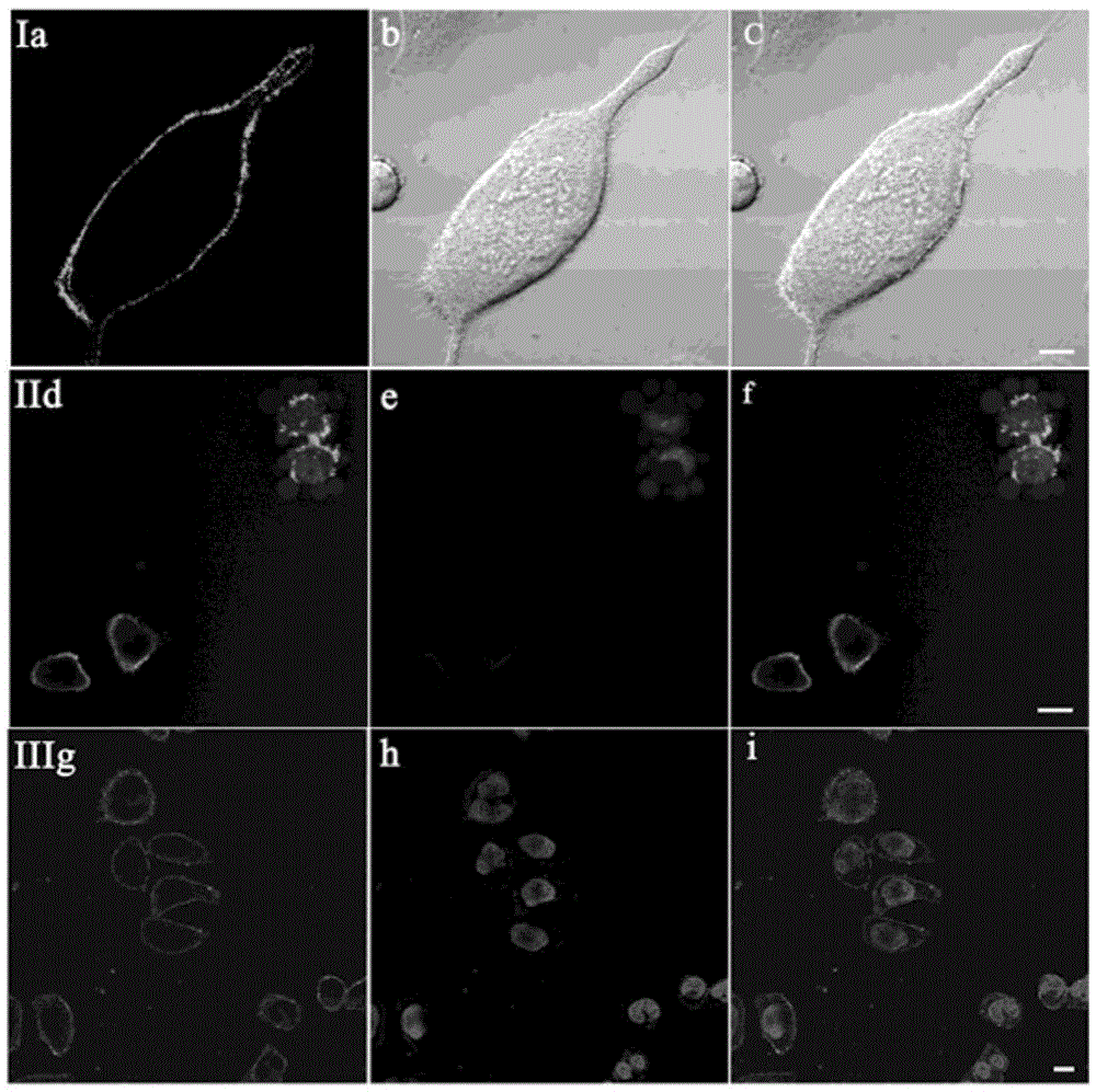
[0039] Stain the attached cell slides with 2 μM T1, and in CO 2 Incubate in the incubator for 20min. After aspirating the culture medium, wash it three times with PBS, take out the cell slide, put the cell growth side down on the glass slide, observe under the two-photon fluorescence microscope, and find that the cell membrane is evenly and continuously stained by T1. Therefore, the probe T1 of the present invention is a two-photon cell membrane probe.

[0040] see results figure 1 (I). Two-photon photographs obtained under 800 nm laser irradiation after staining with T1. Among them, picture a is a two-photon fluorescence photo; picture b is a differential interference picture of bright-field laser scanning; picture c is a superimposed picture of a and b.


PUM


Abstract

The invention discloses a two-photon deep red emission fluorescent probe for imaging cell membranes in tissues based on molecular rotors. The chemical name of the two-photon deep red emission fluorescent probe is N,N-di((4-(2(minute)-(4(second)-dodecyl)pyridine-4(second)iodide)ethylene)phenyl)phenylamine, and a chemical structural general formula is as shown in a formula (I). The invention also discloses application of the two-photon deep red emission fluorescent probe in marking or displaying cell membrane forms in the tissues and in living cells. A test verifies that the two-photon deep red emission fluorescent probe can be used for uniformly and continuously dyeing the cell membranes, bright two-photon deep red light can be emitted after the two-photon deep red emission fluorescent probe is bound to high-viscosity cell membranes because the two-photon deep red emission fluorescent probe is a rotor type molecule, the two-photon deep red emission fluorescent probe is indicated to have a very good prospect by being used as a cell membrane fluorescent probe, and the blank of a cell membrane probe in tissue imaging is hopefully to be filled; meanwhile, the two-photon deep red emission fluorescent probe has the characteristics that the application range is wide, the light stability is good, the cytotoxicity is low, and the cell membranes can be specifically imaged in the living cells.

Description

technical field [0001] The present invention relates to a cell membrane probe with a high signal-to-noise ratio based on a molecular rotor and its application, in particular to a cell membrane fluorescent probe suitable for deep red emission from a near-infrared excitation light source in a two-photon microscope and its use in marking or displaying tissues Cell Membrane Morphology and Applications in Living Cells. Background technique [0002] As the first barrier of eukaryotic cells, the cell membrane ensures the relative stability of the intracellular environment by selectively regulating the entry and exit of certain substances, enabling various biochemical reactions to proceed in an orderly manner. At the same time, the cell membrane is closely related to cell activities such as signal transduction, cell differentiation, cell fusion, and cell recognition. As far as we know, the state and basic functions of many cells can better reflect their real and natural conditions ...

Claims


Application Information

Patent Timeline
Patent Type & Authority Applications(China)
IPC IPC(8): C07D213/38C09K11/06G01N21/64
CPCC07D213/38C09K11/06C09K2211/1007C09K2211/1029G01N21/6486
Inventor 于晓强郭丽方孙渝明田明刚
Owner SHANDONG UNIV
Why Eureka
Try Eureka