Method for quantitatively detecting telomerase activity based on nano pore channel and electrochemical sensing

A nanopore and telomerase technology, applied in the field of biosensing, can solve the problems of poor sensitivity, expensive equipment and reagents, low sensitivity, etc., and achieve the effect of facilitating surface functionalization, mild reaction conditions, and stabilizing the overall structure

Active Publication Date: 2016-07-27
HENAN UNIVERSITY OF TECHNOLOGY
View PDF2 Cites 23 Cited by
  • Summary
  • Abstract
  • Description
  • Claims
  • Application Information

AI Technical Summary

Problems solved by technology

This method is the earliest established method, with good stability and high specificity; the disadvantages are that it requires a large amount of samples, poor sensitivity, long detection time, and is not suitable for the detection of a large number of clinical specimens. The experimenter's body is in danger
TRAP analysis can achieve high-throughput and high-sensitivity detection, but there are still shortcomings caused by PCR amplification technology
In addition, the TRAP method requires the use of expensive equipment and reagents
In addition, TRAP is susceptible to PCR-derived products when screening for potent telomerase inhibitors such as G-tetrad ligands, thus, there are many limitations of this method
[0004] In recent years, in order to replace the traditional TRAP analysis method, researchers have developed many PCR-free analysis methods and applied them to the detection of telomerase activity, such as optical sensors, surface plasmon resonance, electrochemiluminescence, electrochemiluminescence, etc. Chemical detection and exponential isothermal amplification analysis, etc. However, most of the above strategies are limited in application due to the high cost of telomerase primer immobilization, cumbersome operation, and low sensitivity.
Furthermore, most of the current novel telomerase assays are limited to the analysis of telomerase activity in cell extracts and are rarely used in real samples

Method used


Image

  • Method for quantitatively detecting telomerase activity based on nano pore channel and electrochemical sensing
  • Method for quantitatively detecting telomerase activity based on nano pore channel and electrochemical sensing
  • Method for quantitatively detecting telomerase activity based on nano pore channel and electrochemical sensing

Examples


Embodiment 1

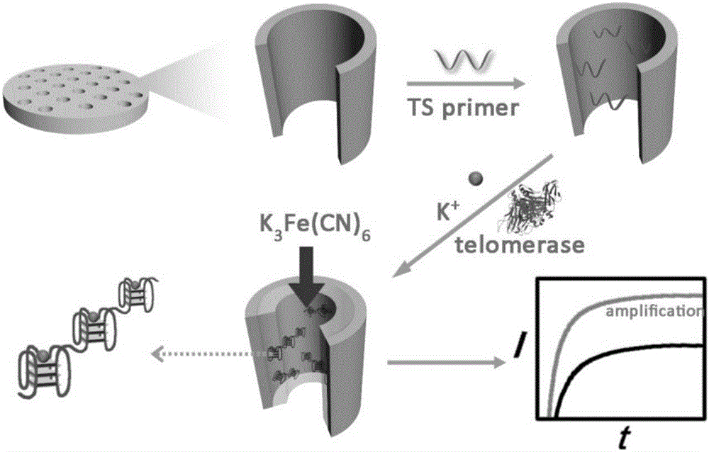
[0046] see figure 1 , an analytical method for detecting telomerase activity in HeLa cells based on nanopores and electrochemical sensing, the detection steps are:

[0047] Step (1) cell culture and telomerase extraction: HeLa cells were inoculated in DMEM medium containing 10% fetal bovine serum, penicillin and streptomycin, and incubated in 5% CO 2 , cultured in a 37°C incubator. All cells were collected during exponential growth phase. Aliquot 1 million cells into 1.5 mL EP tubes, wash twice with ice-cold phosphate buffered saline (pH=7.4), and redisperse in 200 μL of ice-cold CHAPS lysis buffer (10 mM Tris-HCl, pH 7.5 ,1mMMgCl 2 , 1mMEGTA, 0.5% (w / v) CHAPS, 10% (v / v) glycerol, 0.1mMPMSF). Cells were incubated on ice for 30 minutes and then centrifuged at 12000 rpm for 20 minutes at 4°C. After centrifugation, carefully transfer the clarified lysate into a new EP tube, snap freeze and store in a -80°C freezer;

[0048] Step (2) Connect the telomerase primer modified wi...

Embodiment 2

[0054] see figure 1 , an analytical method for detecting telomerase activity in A549 cells, MCF-7 cells and MDA-MB-231 based on nanopores and electrochemical sensing, the detection steps are:

[0055] Step (1) cell culture and telomerase extraction: A549 cells, MCF-7 cells, and MDA-MB-231 cells were inoculated in DMEM medium containing 10% fetal bovine serum, penicillin and streptomycin, and were incubated at 5 %CO 2 , cultured in a 37°C incubator. All cells were collected during exponential growth phase. Aliquot 1 million cells into 1.5 mL EP tubes, wash twice with ice-cold phosphate buffered saline (pH=7.4), and redisperse in 200 μL of ice-cold CHAPS lysis buffer (10 mM Tris-HCl, pH 7.5 ,1mMMgCl 2 , 1mMEGTA, 0.5% (w / v) CHAPS, 10% (v / v) glycerol, 0.1mMPMSF). Cells were incubated on ice for 30 minutes and then centrifuged at 12000 rpm for 20 minutes at 4°C. After centrifugation, carefully transfer the clarified lysate into a new EP tube, snap freeze and store in a -80°C ...

Embodiment 3

[0061] see figure 1 , an analytical method for detecting telomerase activity in A549 cells based on nanopores and electrochemical sensing, the detection steps are:

[0062] Step (1) cell culture and telomerase extraction: A549 cells, MCF-7 cells, and MDA-MB-231 cells were inoculated in DMEM medium containing 10% fetal bovine serum, penicillin and streptomycin, and were incubated at 5 %CO 2 , cultured in a 37°C incubator. All cells were collected during exponential growth phase. Aliquot 1 million cells into 1.5 mL EP tubes, wash twice with ice-cold phosphate buffered saline (pH=7.4), and redisperse in 200 μL of ice-cold CHAPS lysis buffer (10 mM Tris-HCl, pH 7.5 ,1mMMgCl 2 , 1mMEGTA, 0.5% (w / v) CHAPS, 10% (v / v) glycerol, 0.1mMPMSF). Cells were incubated on ice for 30 minutes and then centrifuged at 12000 rpm for 20 minutes at 4°C. After centrifugation, the clarified lysate was carefully transferred to a new EP tube, snap-frozen and stored in a -80 °C freezer. And prepare...


PUM

PropertyMeasurementUnit
pore sizeaaaaaaaaaa

Abstract

The invention discloses a method for quantitatively detecting telomerase activity based on a nano pore channel and electrochemical sensing.The method comprises the following steps that firstly, a telomerase recognition sequence is connected to the inner wall of a porous anodic alumina template or a polyethylene terephthalate film to serve as a primer; secondly, telomerase amplifies the primer to form a G-rich sequence; a G-quadruplex is formed in the presence of potassium ions; electrochemical detection is conducted on the current produced through indicating molecules of the nano pore channel by utilizing an electrochemical workstation.The method does not need complex material preparation and DNA probe marking, and can avoid the defects of high detection cost, complicated operation and poor reproducibility caused by the complex material preparation and DNA probe marking and has the advantages of being low in cost, quick, simple, convenient, high in sensitivity and good in accuracy.

Description

technical field [0001] The invention belongs to a biosensing technology for quantitatively detecting telomerase activity in urine, in particular to a method for quantitatively detecting telomerase activity based on nanopore-based electrochemical sensing. Background technique [0002] Telomerase is an enzyme that synthesizes telomeres at the ends of chromosomes. Telomeres are the natural ends of eukaryotic chromosomes, consisting of thousands of 6-base repeat sequences (TTAGGG), and are essential genetic components of cells. Its function is to maintain the sufficient length of telomeres, ensure the accuracy of genetic information during the replication process, and prevent the loss of genetic information at the end of chromosomes. Under normal circumstances, each cell division cycle will make the telomere shorter and shorter. When the telomere is short enough, it will send a signal and the cell will stop dividing. Telomerase is composed of RNA and protein subunits. It is a ...

Claims


Application Information

Patent Timeline
Patent Type & Authority Applications(China)
IPC IPC(8): G01N27/327
CPCG01N27/3271
Inventor 卫敏刘叙卫伟
Owner HENAN UNIVERSITY OF TECHNOLOGY
Why Eureka
Try Eureka