Intracranial drug eluting stent system and preparation method thereof

A technology of eluting stents and drugs, which is applied in the field of special drug-eluting stent systems, can solve the problems of increased lumen loss in the late stage, reduce restenosis catch-up problems, improve long-term safety, and achieve the effect of vascular function restoration

Active Publication Date: 2016-08-10
赛诺神畅医疗科技有限公司
View PDF4 Cites 8 Cited by
  • Summary
  • Abstract
  • Description
  • Claims
  • Application Information

AI Technical Summary

Problems solved by technology

Significant increases in late lumen loss were demonstrated for both stents by comparing follow-up data at the two time points

Method used


Image

  • Intracranial drug eluting stent system and preparation method thereof
  • Intracranial drug eluting stent system and preparation method thereof
  • Intracranial drug eluting stent system and preparation method thereof

Examples


Embodiment 1

[0051] Example 1 Preparation of intracranial drug-eluting stent

[0052] 1) Preparation of stent base coating

[0053] Use dimethylformamide as a solvent to dissolve n-butyl methacrylate monomer, add sodium nitrate as an electrolyte to increase the conductivity of the solvent, and stir for 30 minutes.

[0054] Table 1. Solution preparation ratio

[0055]

[0056] After the metal stent (the stent material is 316L stainless steel (Germany Euroflex)) was cleaned and dried, it was placed in the reaction vessel containing the above solution, and the polymerization of the stent base coating was completed by applying current and voltage scanning to the solution.

[0057] Electrochemical conditions: voltage: 20V; reaction time: 120 minutes; nitrogen pressure: 2 atmospheres.

[0058] After the reaction, the stent was placed in a vacuum oven to dry.

[0059] 2) Drug coating preparation

[0060] Dissolve polylactic acid polyglycolic acid particles (PLGA, 50 / 50, Mn=90,000-120,000, ...

Embodiment 2

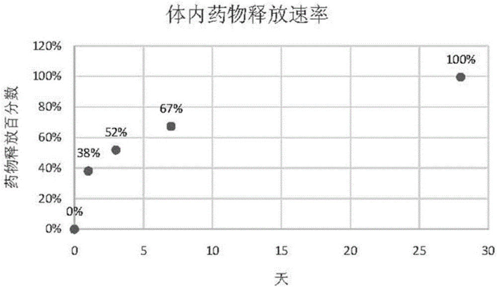
[0070] Embodiment 2 drug release kinetics test

[0071] The drug-eluting stent prepared in Example 1 was implanted in the iliofemoral artery of a rabbit (New Zealand albino rabbit), and the drug concentration of rapamycin in the blood of the rabbit, the concentration of the remaining drug in the stent in the blood vessel, and the concentration of the stent implanted in the artery were tested at different time points. Tissue drug concentration.

[0072] The stent was implanted in the rabbit after anesthesia, and heparin (1000IU / ml, 150IU / Kg) was injected after penetrating the 5F catheter sheath. Insert a 5F balloon catheter into the blood vessel through the catheter sheath to reach the iliac artery, expand it by 10-14 standard atmospheric pressure, and then send the test stent sample into the blood vessel, expand according to the ratio of the balloon size to the blood vessel size 1:1.3, and maintain the pressure After 30 seconds, the balloon delivery system was withdrawn, and ...

Embodiment 3

[0086] Example 3 Intracranial drug-eluting stent coating degradation and endothelial repair test

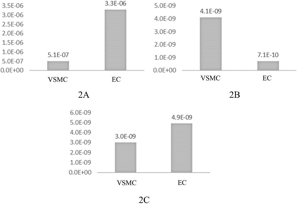
[0087] In a rabbit (New Zealand albino rabbit) iliofemoral artery model, a bare metal stent (Sino Medical) (BMS), a drug-eluting stent (IES) prepared in Example 1 of the present invention, and a base coating without a drug coating were implanted. Layer stent (BS) (its preparation method is identical with embodiment 1, just does not spray drug coating) and another kind of commercially available drug-coated stent (Cypher stent, U.S. Cordis company) (contrast), compare different stent endothelium coverage degree and endothelial activity. Endothelial activity is determined by CD-31 / PECAM-1 staining test, and mature endothelial cells will show a green fluorescent signal after staining.

[0088] The experimental results showed that after 90 days of implantation, both IES and BS achieved complete coverage of the endothelium, which was comparable to the BMS stent, and there was no drug ...


PUM

PropertyMeasurementUnit
thicknessaaaaaaaaaa

Abstract

The invention relates to an intracranial drug eluting stent system and a preparation method thereof, in particular to an intracranial drug eluting stent used for treating the intracranial atherosclerotic stenosis disease .The intracranial drug eluting stent is composed of a metal stent body and a coating structure covering the surface of the metal stent body, the coating structure comprises one or more stent substrate coatings and drug coatings, and the drug coatings contain a biodegradable drug carrier and a drug inhibiting excessive proliferation of VSMCs .According to the intracranial drug eluting stent, the diseased artery is expanded with the stent to improve intracranial artery blood perfusion, the drug carried by the stent can prevent excessive proliferation of endangium, and thus the probability of in-stent restenosis is lowered; meanwhile, stent healing in the artery can be rapidly achieved, and thus long-term safety and effectiveness are ensured for patients.

Description

technical field [0001] The invention relates to a special drug-eluting stent system for treating intracranial artery stenosis. Specifically, the present invention relates to an intracranial drug-eluting stent for treating intracranial atherosclerotic stenosis. The stent expands diseased blood vessels and improves intracranial arterial blood perfusion. The drugs contained in the stent can prevent blood vessel Hyperplasia of the intima reduces the probability of restenosis in the stent, and the stent can quickly heal in the artery, thereby ensuring the long-term safety and effectiveness of the patient. Background technique [0002] Stroke is a serious global health problem, it is the third leading cause of death in the world after cancer and myocardial infarction, and 3% of adult disability is caused by stroke. Atherosclerotic intracranial artery stenosis is an important cause of ischemic stroke. The WASID (Warfarin-Aspirin Study of Symptomatic Intracranial Arterial Stenosis...

Claims


Application Information

Patent Timeline
Patent Type & Authority Applications(China)
IPC IPC(8): A61L31/02A61L31/10A61L31/08A61L31/16
CPCA61L31/022A61L31/08A61L31/10A61L31/16A61L2300/216A61L2300/416A61L2420/06A61L2420/08C08L67/04
Inventor 孙箭华康小然曹懿舜李天竹吴祥芬
Owner 赛诺神畅医疗科技有限公司
Why Eureka
Try Eureka