Preparation method of surface-mineralized chitosan microsphere

A technology of chitosan microspheres and chitosan, applied in coatings, medical science, prostheses, etc., can solve the problem of insignificant effects, inability to obtain components and structures, and the growth of nano-hydroxyapatite cannot reach natural bone. Issues such as the required content of inorganic minerals to achieve the effect of large-scale production, good biological activity and biological function, and good osteogenic activity

Inactive Publication Date: 2016-10-26
TAIYUAN UNIV OF TECH
View PDF2 Cites 4 Cited by
  • Summary
  • Abstract
  • Description
  • Claims
  • Application Information

AI Technical Summary

Problems solved by technology

[0004] Some scholars have successfully synthesized nano-hydroxyapatite/chitosan composite materials by using the biomimetic mineralization method. It also has some characteristics similar to natural bone in structure, but the effect is no

Method used


Image

  • Preparation method of surface-mineralized chitosan microsphere
  • Preparation method of surface-mineralized chitosan microsphere

Examples


Embodiment 1

[0022] Mix 110 g of liquid paraffin with 3 g of span80, slowly add 50 g of 2 wt% chitosan acetic acid solution after stirring, stir and emulsify for 3 h at 30 °C to form a water-in-oil emulsion.

[0023] Add 0.0428 g of glutaraldehyde dropwise for cross-linking for 3 h to obtain a uniformly mixed emulsion, which is left to settle;

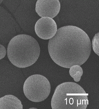
[0024] Add petroleum ether and isopropanol to wash the precipitate, and dry at 50-60°C for 10 h to obtain chitosan microspheres with a particle size of 5-15 μm (see figure 1 ).

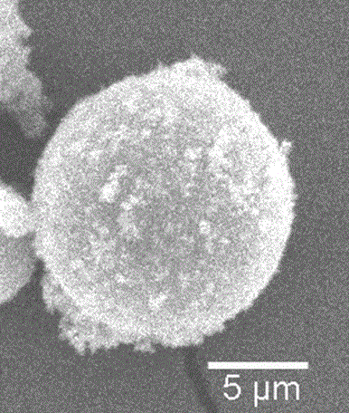
[0025] 0.3 g of chitosan microspheres were fully dispersed in 250 mL of mineralization solution with a pH of 4.5, and the reaction was continuously stirred at 37 °C for 3 h to obtain a uniformly mixed solution. Set aside to settle, wash the precipitate with deionized water and isopropanol successively, and dry to obtain the mineralized chitosan microspheres. The surface of the mineralized chitosan microspheres has an acicular nano-apatite structure (see figure 2 ).

Embodiment 2

[0027] 124.742 g of liquid paraffin and 3.858 g of span80 were mixed, and after stirring, 60 g of 4 wt% chitosan acetic acid solution was slowly added, stirred and emulsified at 37°C for 3 h to form a water-in-oil emulsion.

[0028] 0.214 g of glutaraldehyde was added dropwise for cross-linking for 3 h, and a uniformly mixed emulsion was obtained. Let the precipitate stand still, add petroleum ether and isopropanol to wash the precipitate, and dry at 50-60°C for 12 h.

[0029] 0.4 g of chitosan microspheres were fully dispersed in 250 mL of mineralization solution with a pH of 5, and the reaction was continuously stirred at 70 °C for 12 h to obtain a uniformly mixed solution. Let the precipitate stand, wash the precipitate with deionized water and isopropanol in turn, and dry to obtain mineralized chitosan microspheres.

Embodiment 3

[0031] 124.742 g of liquid paraffin and 3.858 g of span80 were mixed, and after stirring, 50 g of 4 wt% chitosan acetic acid solution was slowly added, and stirred and emulsified at 60°C for 3 h to form a water-in-oil emulsion.

[0032] 0.214 g of vanillin was added dropwise for cross-linking for 3 h, and a uniformly mixed emulsion was obtained. Let the precipitate stand still, add petroleum ether and isopropanol to wash the precipitate, and dry at 50-60°C for 11 h.

[0033] 0.35 g of chitosan microsphere powder was fully dispersed in 250 mL of mineralization solution with a pH of 5.5, and the reaction was continuously stirred at 85 °C for 12 h to obtain a uniformly mixed solution. Let the precipitation stand still, wash the precipitation with deionized water and isopropanol in turn, and dry to obtain the mineralized chitosan microspheres.


PUM


Abstract

The invention relates to a preparation method of a surface-mineralized chitosan microsphere and belongs to the field of medical materials. The preparation method comprises the following steps: (1) preparing a chitosan microsphere by utilizing an emulsion-crosslinking method; (2) configuring a mineralized solution; (3) putting prepared chitosan microsphere powder into the configured mineralized solution to enable mineralization reaction for a certain time at a certain temperature, standing and precipitating, washing and drying to obtain a chitosan microsphere surface-mineralized composite material. A large number of uniformly-dispersed lamellar nano hydroxyapatite crystals are generated on the surface of the chitosan microsphere in situ through a mineralization method. According to the preparation method provided by the invention, under mild conditions, the accurate control of the components, the structure and the content of the nano hydroxyapatite crystals on the surface of the polymer microsphere are realized, so that a bone-like apatite surface with high surface activity and good osteogenetic activity is prepared; the preparation method is stable in process and low in cost and can realize large-scale production. The preparation method provided by the invention has a wide application value in clinical aspect of bone tissue engineering.

Description

technical field [0001] The invention relates to a preparation method for surface mineralization of chitosan microspheres, which belongs to medical biomaterials and has wide application significance in the clinical aspect of bone tissue engineering. Background technique [0002] Nano-hydroxyapatite is the main component of human bone inorganic matter, has excellent physical and chemical properties and biological effects, and is an ideal substitute material for bone repair. However, nano-hydroxyapatite prepared by traditional methods has disadvantages such as large particles, uneven dispersion, agglomeration, and weak bonding strength with the matrix, and there are also high cost, difficult control, and poor repeatability in the preparation method. insufficient. The mineralization method is to use the biomineralization mechanism to regulate the nucleation and growth of inorganic nanocrystals through electrostatic matching or chemical bonding, so that organic substances can co...

Claims


Application Information

Patent Timeline
IPC IPC(8): C08J3/24C08J3/12C08J7/00C08J7/06C08L5/08A61L27/12A61L27/20A61L27/50
CPCC08J3/24A61L27/12A61L27/20A61L27/50A61L2430/02C08J3/12C08J7/00C08J7/06C08J2305/08
Inventor 黄棣胡银春王楷群杜晶晶牛璐璐魏延连小洁
Owner TAIYUAN UNIV OF TECH
Why Eureka
Try Eureka