
Method for screening melanin formation resistant medicines on basis of magnetic bead separation

An anti-melanin, magnetic bead separation technology, applied in material separation, analytical materials, measurement devices, etc., can solve the problems of unreported, time-consuming, changing the binding properties of proteins and compounds, and achieve reasonable process design and good reproducibility. , the effect of shortening the screening time

Inactive Publication Date: 2017-01-04
NANJING UNIVERSITY OF TRADITIONAL CHINESE MEDICINE
View PDF4 Cites 3 Cited by
  • Summary
  • Abstract
  • Description
  • Claims
  • Application Information

AI Technical Summary

Problems solved by technology

Of course, there are certain limitations in the use of magnetic beads for drug screening. First, the covalent binding of proteins to magnetic beads may change the binding properties of proteins and compounds. Second, the automated process of this method is quite time-consuming. Optimizing multiple factors such as diameter, pressure, protein-magnetic bead ratio, etc.
At present, no one has reported the method of selecting tyrosinase as a drug target and establishing a method for screening tyrosinase inhibitors based on magnetic separation by applying magnetic bead bonding technology.

Method used


Image

  • Method for screening melanin formation resistant medicines on basis of magnetic bead separation
  • Method for screening melanin formation resistant medicines on basis of magnetic bead separation
  • Method for screening melanin formation resistant medicines on basis of magnetic bead separation

Examples


Embodiment 1

[0028] Example 1 Preparation of tyrosinase-bonded magnetic beads

[0029] Take a certain volume of magnetic beads, use an equal volume of 25mM MES (pH 6) solution to wash twice, twice for 10min, add an equal volume of freshly prepared EDC solution and NHS solution (50mg / ml) to the washed magnetic beads, Mix well and incubate with slow shaking for 30 min at room temperature. After incubation, put the tube on the magnet for 4 minutes, remove the supernatant, and finally wash twice with 25mM MES (pH6) solution, and set aside.

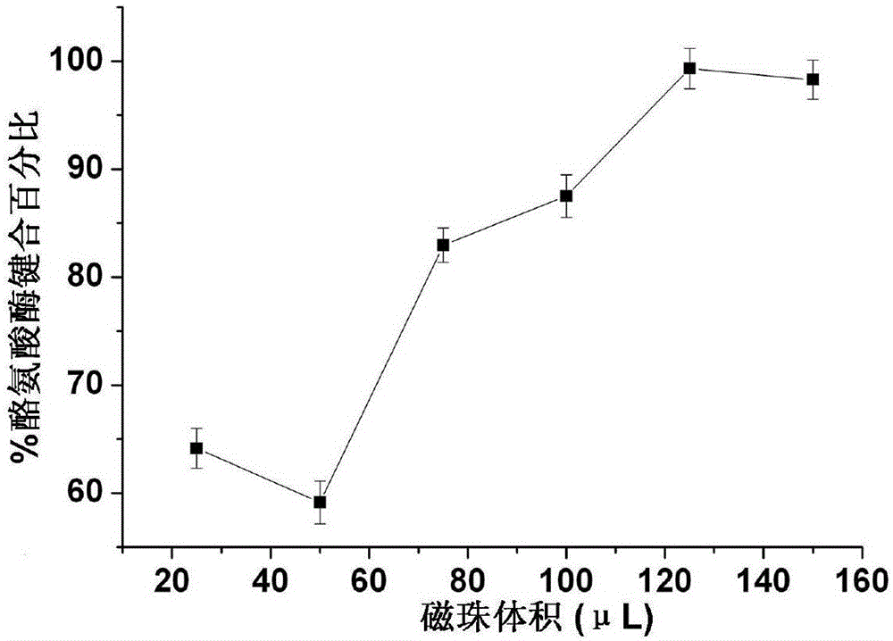
[0030] Take 25, 50, 75, 100, 125, and 150 μL of activated magnetic beads respectively, add 100 μL of tyrosinase (1 mg / ml) solution, shake and mix well, and incubate overnight at room temperature. After incubation, place the tube on the magnetic beads for 4 minutes, remove the supernatant remaining from the binding, and use the Bradford method to determine the protein content in it. Add 100 μL of 0.5% BSA solution to the bonded magnetic beads and shake fo...

Embodiment 2

[0036] Example 2 Research on various factors of the drug screening method for anti-melanin production based on magnetic bead separation

[0037] (1) Using paeoniflorin as a model drug, optimize the denaturation and elution solvent

[0038] Take 20 μL of tyrosinase-bonded magnetic beads prepared in Example 1 above, add 20 μL of paeoniflorin reference solution (0.1 mg / ml) and 160 μL of PBS buffer solution (pH7.4) and mix, and incubate for 30 min. After the incubation is completed, Wash 4 times with 200 μL PBS buffer solution, and the eluate enters the liquid phase for analysis. Finally, the volume concentration is 10%, 30%, 50%, 70% and 90% acetonitrile-water solution (v / v) and the volume concentration is 10%. , 30%, 50%, 70% and 90% methanol-water solution (v / v) for denaturation elution, and the denaturation eluate was taken for liquid phase analysis.

[0039] (2) Using paeoniflorin as a model drug, optimize the incubation time

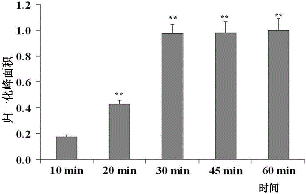
[0040] Take 20 μL of tyrosinase-bonded magneti...

Embodiment 3

[0053] Example 3 Screening of Anti-Melanin Production Drugs in Sanbai Decoction Components

[0054] (1) Take a certain volume of magnetic beads, wash them twice with an equal volume of 25mM MES buffer solution, each time for 10min, after washing, add an equal volume of 50mg / ml EDC solution and 50mg / ml NHS solution, mix well, and store at room temperature Incubate with slow shaking, after incubation, place on the magnet for 4min, remove the supernatant, and then wash with 25mM MES buffer solution to obtain activated magnetic beads, set aside;

[0055] (2) Take 125 μl of the activated magnetic beads obtained in step (1), add 100 μg of tyrosinase, shake and mix evenly, incubate at room temperature, after incubation, place on the magnetic beads for 4 minutes, and remove the remaining supernatant for bonding , to obtain tyrosinase-bonded magnetic beads;

[0056] (3) Weigh 5g Radix Paeoniae Alba, 5g Atractylodes Rhizoma Atractylodes Rhizoma Atractylodes Rhizoma Atractylodes Rhizoma...


PUM


Abstract

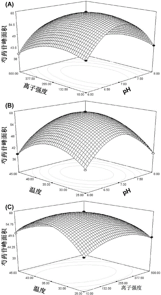
The invention discloses a method for screening melanin formation resistant medicines on the basis of magnetic bead separation. Target protein conjugates are analyzed by the aid of magnetic separation and chemical information acquisition (liquid chromatography-mass spectrometry) technologies, and the method can be used for screening and evaluating melanin formation resisting effects of the medicines. The optimal incubation temperatures, the optimal incubation time, the optimal pH (potential of hydrogen) values of solution during incubation, the optimal ion strength and the optimal denaturation cleaning solution are screened by the aid of large quantities of experiments. Compared with the traditional methods for screening melanin formation resistant medicines, the method has the advantages that the screening efficiency can be obviously improved, and the screening time can be shortened; synergistic effects among compounds can be discovered, the method can be used for screening large-scale natural product libraries or combinatory chemical libraries, is suitable for screening the melanin formation resistant medicines (including chemically synthetic medicines and natural products) and discovering lead compounds at early stages, is speedy and accurate and is good in repeatability, and the like.

Description

technical field [0001] The invention relates to a drug screening method, in particular to a drug screening method for anti-melanin production based on magnetic bead separation with high screening efficiency. Background technique [0002] The black and white of the skin mainly depends on the ability of melanocytes to synthesize melanin. Among the basal cells of the human epidermis, there are melanocytes, which contain tyrosinase that can oxidize tyrosine into polysaccharides, and then go through a series of metabolic processes in the middle to finally produce melanin. The more melanin produced, the darker the skin; otherwise, the fairer the skin. [0003] Tyrosinase has a unique dual catalytic function and is a key enzyme in the synthesis of melanin in organisms, which is closely related to human aging. Its abnormal overexpression can lead to pigmentation diseases in human body. Tyrosinase inhibitors can treat current common pigmented skin diseases such as freckles, chloas...

Claims


Application Information

Patent Timeline
Patent Type & Authority Applications(China)
IPC IPC(8): G01N30/02
CPCG01N30/02G01N2030/027
Inventor 陶益苏丹丹杜映姗蔡宝昌
Owner NANJING UNIVERSITY OF TRADITIONAL CHINESE MEDICINE
Learn More