Electrochemical sensor for quick detection of plasma indole
An electrochemical and sensor technology, applied in the field of electrochemical detection, achieves the effects of simple operation, simple operation, simple production and modification process
- Summary
- Abstract
- Description
- Claims
- Application Information
AI Technical Summary
Problems solved by technology
Method used
Image
Examples
Embodiment 1
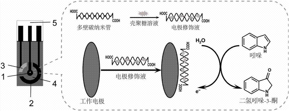
[0037] Embodiment 1: the preparation method of the electrochemical sensor that detects plasma indole
[0038] 1) Prepare the screen printing electrode of the present invention:
[0039] The screen printing electrode of the present invention prints carbon paste, silver / silver chloride paste and insulating paste sequentially on a polyethylene terephthalate (PET) substrate. Specifically include the following steps:
[0040] ①Clean the PET substrate, print carbon paste on the PET substrate after drying, make working electrodes and auxiliary electrodes, and dry at room temperature;
[0041] ②Print silver paste containing silver chloride on the above-mentioned PET substrate to make a reference electrode and dry at room temperature;
[0042] ③Avoid the circular working area, print insulating paste on the above PET substrate to cover the wires;
[0043] ④The above-mentioned working electrode, auxiliary electrode and reference electrode form a circular working area, and each electro...
Embodiment 2
[0051] Embodiment 2: the use method of the electrochemical sensor that detects plasma indole
[0052]1) Purchasing analytically pure ether for pretreatment of plasma samples;
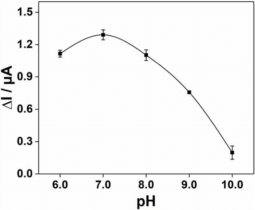
[0053] 2) Prepare phosphate buffer: prepare 0.1mol L respectively -1 Sodium dihydrogen phosphate and disodium hydrogen phosphate solution are mixed according to a certain volume ratio to prepare a pH=7.0 phosphate buffer solution, which is placed in a container;
[0054] 3) Take 300 μL of plasma sample and 600 μL of the ether solution in a test tube, shake on a shaker at 250 rpm for 18 minutes, then centrifuge at 13300 g at 4°C for 5 minutes, and finally take the supernatant and dry it with nitrogen at 30°C;
[0055] 4) Reconstitute the treated sample with 150 μL ultrapure water;
[0056] 5) Take equal volumes of the solution and phosphate buffer solution obtained in step 3) respectively, vortex and mix to obtain a reaction mixture;
[0057] 6) Connect the screen printing electrode to CHI852C electro...
Embodiment 3
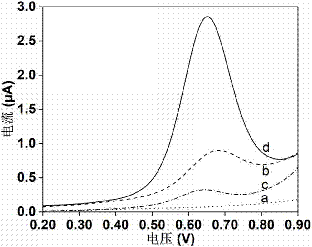
[0059] This example is to investigate the electrochemical response to indole before and after the screen printing electrode modification. Use the bare electrode and the modified electrode to measure the indole standard solution respectively, the results are shown in figure 2 . The dotted line a and the short horizontal line b in the figure represent the measurement of phosphate buffer and 100.0 μg L -1 The voltammetric curve of the indole standard, the dotted line c and the solid line d in the figure respectively represent the determination of phosphate buffer and 100.0 μg L by the modified electrode -1 Voltammetric curves of indole standards. The results showed that the obtained electrochemical signal was produced by the oxidation of indole. In addition, the electrochemical response signal of indole measured by the modified electrode was significantly improved, and the peak shape of the voltammetry curve was obviously improved.
PUM
 Abstract
Description
Claims
Application Information
 - R&D
- Intellectual Property
- Life Sciences
- Materials
- Tech Scout
- Unparalleled Data Quality
- Higher Quality Content
- 60% Fewer Hallucinations
Browse by: Latest US Patents, China's latest patents, Technical Efficacy Thesaurus, Application Domain, Technology Topic, Popular Technical Reports.
© 2025 PatSnap. All rights reserved.Legal|Privacy policy|Modern Slavery Act Transparency Statement|Sitemap|About US| Contact US: help@patsnap.com


