

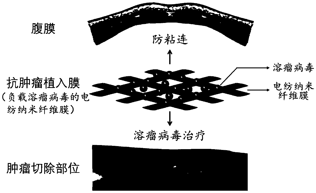
A kind of anti-tumor implant film and preparation method thereof
An anti-tumor and oncolytic technology, which is applied in the field of anti-tumor implantation membrane and its preparation, can solve the problems that are difficult to realize, and achieve the effects of avoiding toxic and side effects, reducing drug usage, and increasing drug concentration
- Summary
- Abstract
- Description
- Claims
- Application Information
AI Technical Summary
Problems solved by technology
Method used
Image

Examples
Embodiment 1
[0033] Example 1: Preparation method of electrospun nanofibrous membrane loaded with oncolytic virus
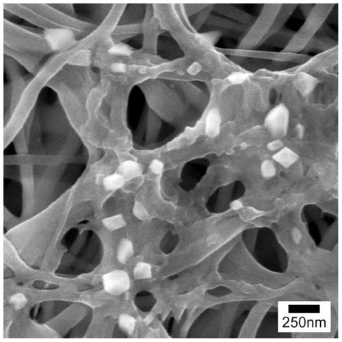
[0034] Chitosan (viscosity-average molecular weight is about 850,000, deacetylation degree is 95%) and polyethylene oxide (weight-average molecular weight is 600,000) are dissolved in 90% acetic acid solution together, and the mass fraction is configured as 3.25%. Polymer solution, wherein the mass ratio of chitosan to polyethylene oxide is 90:10. The polymer solution was placed in the sampling device of the electrospinning device, the voltage was adjusted to 20kv, the distance between the nozzle and the collecting plate was 15cm, and the nanofiber film with a thickness of 150-200μm was collected. Remove the nanofibrous membrane from the collection plate, soak it in 1mol / L potassium carbonate solution for 1hr, then wash it with deionized water and dry it at room temperature to remove the unvolatile acetic acid solution and polyethylene oxide, and release chitosan positively ...
Embodiment 2
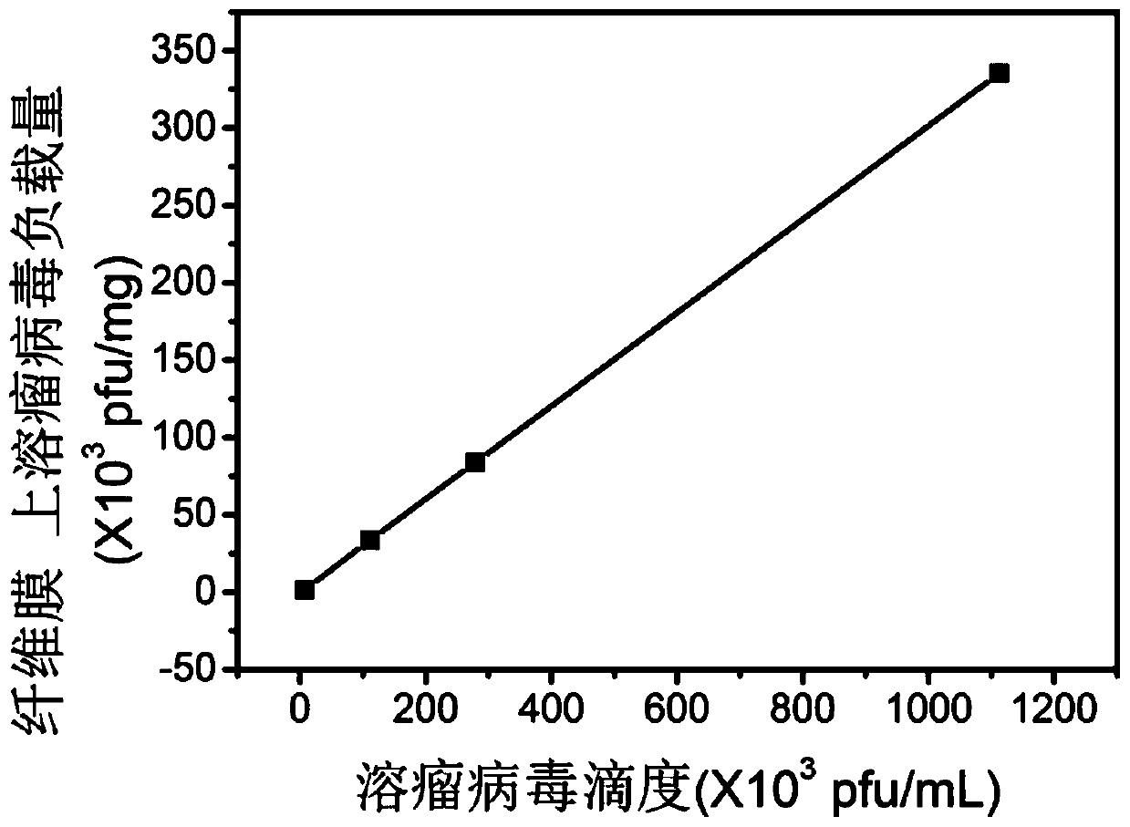
[0037] Example 2: Control of Oncolytic Virus Load
[0038] Divide the area to 1×1cm 2 , an electrospun nanofibrous membrane weighing 1.5 mg was soaked in 0.5 mL of oncolytic virus solutions with different virus titers, and stood at 4 °C for 2 hr. The change of virus titer in the oncolytic virus solution before and after the bubble membrane was detected, and the difference was the virus load loaded on the electrospun nanofiber membrane.
[0039] Such as figure 2 As shown in the curve, when the concentration of virus in the oncolytic virus solution before the bubble membrane is from 7×10 3 , 1.11×10 5 , 2.79×10 5 increased to 1.11×10 6 pfu / mL, the load of oncolytic virus loaded on the electrospun nanofiber membrane then changed from 1.29×10 3 , 3.35×10 4 , 8.4×10 4 Increased to 3.35×10 5 pfu / mg, that is, the greater the concentration of virus in the oncolytic virus solution before the bubble membrane, the more oncolytic virus loaded onto the electrospun nanofiber membr...
Embodiment 3
[0040] Example 3: Tumor therapeutic effect of electrospun nanofibrous membrane loaded with oncolytic virus
[0041] 1) Cell culture
[0042] Human liver cancer cells Bel-7402 (referred to as 2D-7402) were filled with DMEM complete medium containing 10% FBS and 1% double antibody, placed in 5% CO 2 , cultured and subcultured in a constant temperature incubator at 37°C. The growth was observed with an inverted microscope, and the cells were passaged every 2 days, and the cells in the logarithmic growth phase were taken for cytotoxicity experiments.
[0043] The Bel-7402 drug-resistant cell line 7402-5fu resistant to chemotherapy drug 5-fluorouracil (5-fu) is cultivated in a similar way to 2D-7402, except that its culture medium is DMEM complete medium containing 20 μg / mL 5-fu.
[0044] Bel-7402 cells cultured with 3D cell culture technology have the properties of stem cells, which are recorded as 3D-7402.
[0045] The culture method of human liver cancer cell HepG2 is the sam...
PUM
| Property | Measurement | Unit |
|---|---|---|
| diameter | aaaaa | aaaaa |
| diameter | aaaaa | aaaaa |
Abstract
Description
Claims
Application Information