Fluorescent carbon dot for labeling of radionuclide iodine, synthetic method and application

A technology of radionuclide and fluorescent carbon dots, which is applied in the fields of nanomedicine, molecular imaging, and nuclear medicine, can solve problems such as complicated operation and difficult purification, and achieve high fluorescence yield, good physical stability and radiochemical stability, The effect of strong radiochemical stability

Active Publication Date: 2017-09-01
XIAMEN UNIV
View PDF1 Cites 4 Cited by
  • Summary
  • Abstract
  • Description
  • Claims
  • Application Information

AI Technical Summary

Problems solved by technology

For different target molecules, it is usually necessary to select different labeling methods according to the structure and biochemical properties of the labeled molecule, which is not only complicated to operate, but also difficult to purify

Method used


Image

  • Fluorescent carbon dot for labeling of radionuclide iodine, synthetic method and application
  • Fluorescent carbon dot for labeling of radionuclide iodine, synthetic method and application
  • Fluorescent carbon dot for labeling of radionuclide iodine, synthetic method and application

Examples


Embodiment 1

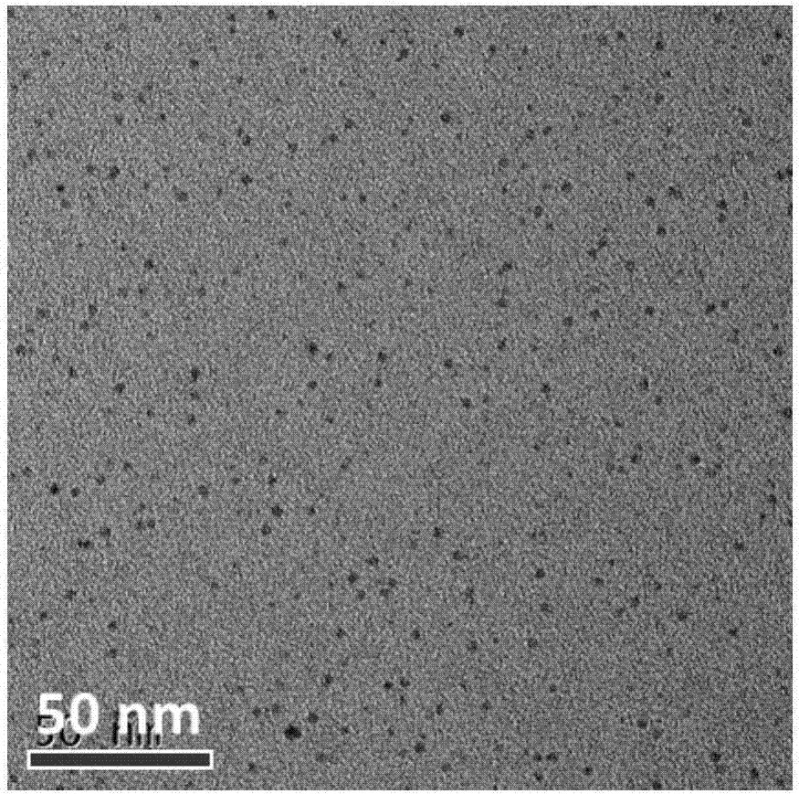
[0025] Dissolve 0.5g of citric acid and 0.1g of tyrosine in water, adjust the pH to 1, make the solution clear, stir evenly, put it in a high-pressure reactor, and react at 180°C for 6 hours. After cooling, put The resulting carbon dot solution was purified by dialysis for 24 h with a molecular weight 500 dialysis bag.

[0026] Dissolve 10 mg of carbon dots in DMF, add 20 mg of EDC and 20 mg of NHS for activation for two hours, then add 100 mg of methoxy PEG2000 amino, react overnight, and dialyze with a dialysis bag with a molecular weight of 2000 for one day.

[0027] Take 10 μg of PEG-modified carbon dots (dissolved in 100 μl of water) into the EP tube coated with 100 μg of chloroglycoside, and then add 1 mCi of Na 125 I solution, shaking and reacting for half an hour, take out the solution to obtain the nuclide 125 I labeled carbon dots.

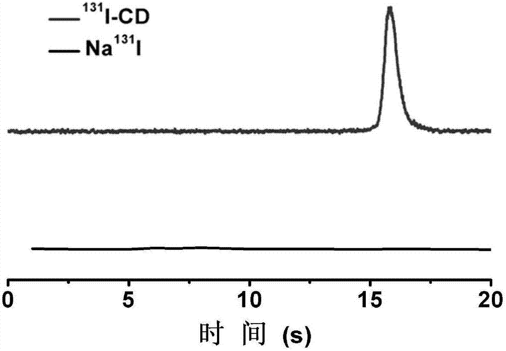
[0028] The TEM image of fluorescent carbon dots, the fluorescence spectrum image of fluorescent carbon dots, the HPLC image of carbon...

Embodiment 2

[0030] Dissolve 0.6g citric acid and 0.3g tyrosine in water, adjust pH=1.5, make the solution clear, stir evenly, place in a high-pressure reactor, react at 160°C for 12h, after cooling, put The obtained carbon dot solution was purified by silica gel column.

[0031] Dissolve 15 mg of carbon dots in DMF, add 30 mg of EDC and 30 mg of NHS for activation for two hours, then add 150 mg of folic acid-PEG2000-amino, react overnight in the dark, and then dialyze with a dialysis bag with a molecular weight of 2000 for two days.

[0032] Take 20 μg of folic acid-modified carbon dots (dissolved in 100 μl of water) and add to an EP tube containing 200 μg of chloramine T, and then add 1 mCi of Na 131 I solution, shaking and reacting for 10 minutes, take out the solution to obtain the nuclide 131 I labeled carbon dots.

Embodiment 3

[0034] Dissolve 0.4g citric acid and 0.4g tyrosine in water, adjust the pH=1.5, make the solution clear, stir evenly, put it in a high-pressure reactor, and react at 200°C for 4 hours. After cooling, put The resulting carbon dot solution was purified by dialysis for 24 h with a molecular weight 500 dialysis bag.

[0035] Dissolve 12 mg of carbon dots in DMF, add 25 mg of EDC and 25 mg of NHS for activation for two hours, then add 10 mg of c(RGDfk) polypeptide, react overnight in the dark, and dialyze with a dialysis bag with a molecular weight of 1000 for two days.

[0036] Take 15 μg of RGD-modified carbon dots (dissolved in 120 μl of water) and add them to the EP tube coated with 150 μg of chloroglycoside, and then add 1 mCi of Na 124 I solution, shaking and reacting for 1 hour, take out the solution to obtain the nuclide 124 I labeled carbon dots.


PUM


Abstract

The invention belongs to the field of nano-medicine and molecular imaging, and particularly relates to a fluorescent carbon dot for labeling of radionuclide iodine, a synthetic method of the fluorescent carbon dot and application in tumor imaging. The carbon dot is synthesized in one step and can be directly labeled with radionuclide iodine. A labeled product has excellent radiochemical stability and physicochemical stability and can be directly used for 124I-based PET (positron emission tomography), SPECT(125I-based single-photon emission computed tomography) imaging and 131I radiotherapy of a tumor region. The synthetic method of the fluorescent carbon dot is simple, price of raw materials is low, fluorescence efficiency is high, and biocompatibility is good; the fluorescent carbon dot has the advantage of rapid metabolism in vivo like small molecules, and can also be well enriched in the tumor region by effects of high permeability and long retention of nanoparticles to diagnose and treat a tumor.

Description

technical field [0001] The invention belongs to the fields of nanomedicine, molecular imaging and nuclear medicine, and in particular relates to a fluorescent carbon dot which can be directly used for radionuclide iodine labeling, a synthesis method thereof, and an application of tumor imaging. Background technique [0002] Nanomedicine is a new type of science and technology that uses nanotechnology to carry out medical applications such as disease prevention, diagnosis and treatment, and health improvement at the molecular level. Due to the small size, large specific surface area, precisely adjustable morphology and unique physical and chemical properties of nanomaterials, they are widely used in many fields of biology and medicine (such as bioluminescent labeling, in vivo multimodal imaging, pharmaceuticals, etc.) and gene transport, tumor multifunctional therapy, etc.), all have broad application prospects. [0003] As a new type of ultra-small carbon nanoparticles, flu...

Claims


Application Information

Patent Timeline
Patent Type & Authority Applications(China)
IPC IPC(8): C09K11/65A61K51/02C09K11/06A61K51/04
CPCA61K51/02A61K51/0406C09K11/06C09K11/65C09K2211/1007
Inventor 孙晓莲刘念宋梦麟孙文静
Owner XIAMEN UNIV
Why Eureka
Try Eureka