Whole eye cornea bioengineering cultivation method

A technology of cornea and culture method, which is applied in the direction of biochemical equipment and methods, microorganisms, cell culture support/coating, etc., and can solve the problems of increasing infection, transfection, operation failure, etc.

Inactive Publication Date: 2018-07-13
山东麦德克斯生物科技有限公司
View PDF10 Cites 4 Cited by
  • Summary
  • Abstract
  • Description
  • Claims
  • Application Information

AI Technical Summary

Problems solved by technology

[0005] Other tissue engineering techniques use heterogeneous or allogeneic cells as seed cells to cultivate full-thickness biological corneas on scaffolds, whi

Method used


Image

  • Whole eye cornea bioengineering cultivation method
  • Whole eye cornea bioengineering cultivation method
  • Whole eye cornea bioengineering cultivation method

Examples


Embodiment 1

[0098] Corneal epidermal culture

[0099] The patient’s lidocaine was locally anesthetized, and the patient’s oral cavity was locally disinfected with povidone iodine. The patient’s oral mucosal tissue was taken from the internal buccal mucosal epithelium, about 3×3mm in size.

[0100] Treat the oral mucosal tissue with 5ml dispase II (3mg / ml, Roche) at 37℃ for about 1 hour. After removing all the epithelial layer, the oral mucosal epithelial cells are collected and placed in the digestive juice for 15 minutes to form a single cell suspension liquid.

[0101] The separated cells are put into a collagen medium, the surface of the medium is amniotic membrane, and the amniotic membrane is coated with N-isopropyl polymer.

[0102] At the same time, T3T feeder cells and mitomycin C are placed in the periphery to secrete growth hormone and cytokines.

[0103] After culturing at 37°C for about 2 weeks, an active 3-5 layer of epidermal cells can be obtained by the naked eye. After hematoxylin-...

Embodiment 2

[0106] Corneal stroma culture

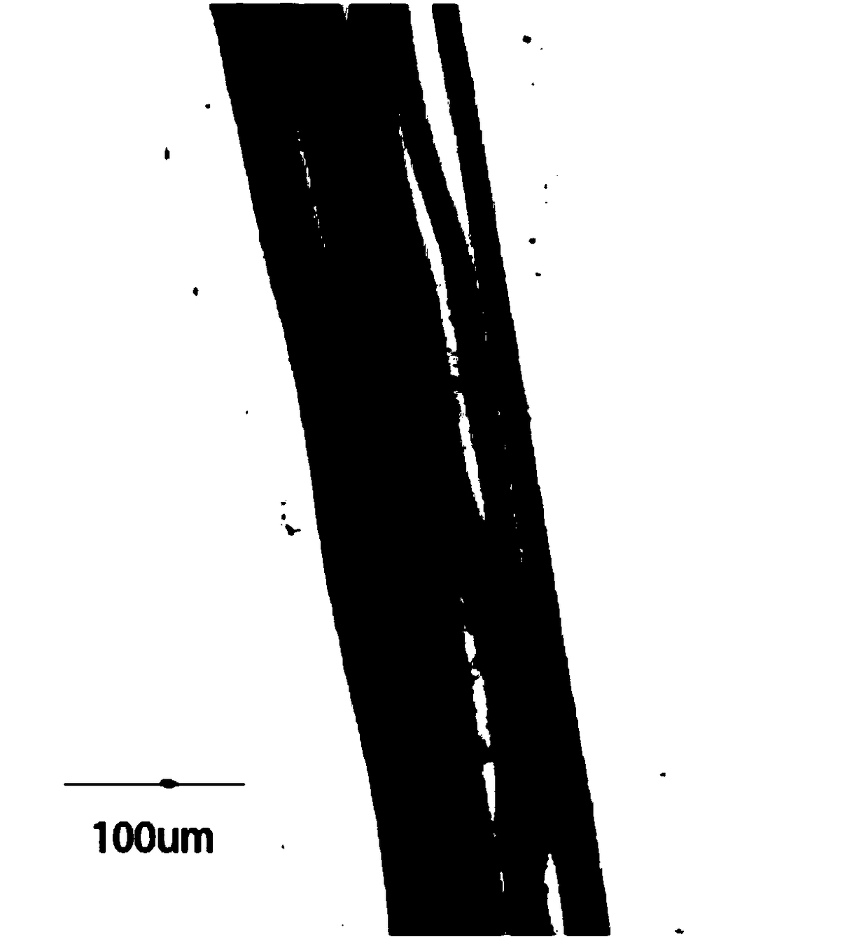
[0107] 1. Isolation of autologous skin fibroblasts

[0108] Under aseptic conditions, remove the 3×2mm size of the skin tissue of the inner thigh of the lower limbs and place it in phosphate buffered saline (PBS) containing 100U / ml penicillin and 100ug / ml streptomycin to wash 3-5 times to remove blood stains and Cut off the connective tissue.

[0109] The skin is soaked in 10ml of a mixture containing 2ml of double-antagonist (100U / ml penicillin and 100μg / ml streptomycin) and 1ml of digestive solution, kept at 4°C, and left overnight.

[0110] The next day, continue to use phosphate-buffered saline containing penicillin and streptomycin (double antibodies) to wash 3-5 times, and then separate the dermis and epidermis, use a new sterile scalpel to cut the dermis into small pieces. The fragments were transferred to a 50ml centrifuge tube containing 0.5ml of biantagonist and 5ml of collagenase (1mg / ml).

[0111] Incubate at 37°C for 4-8 hours, shaking appr...

Embodiment 3

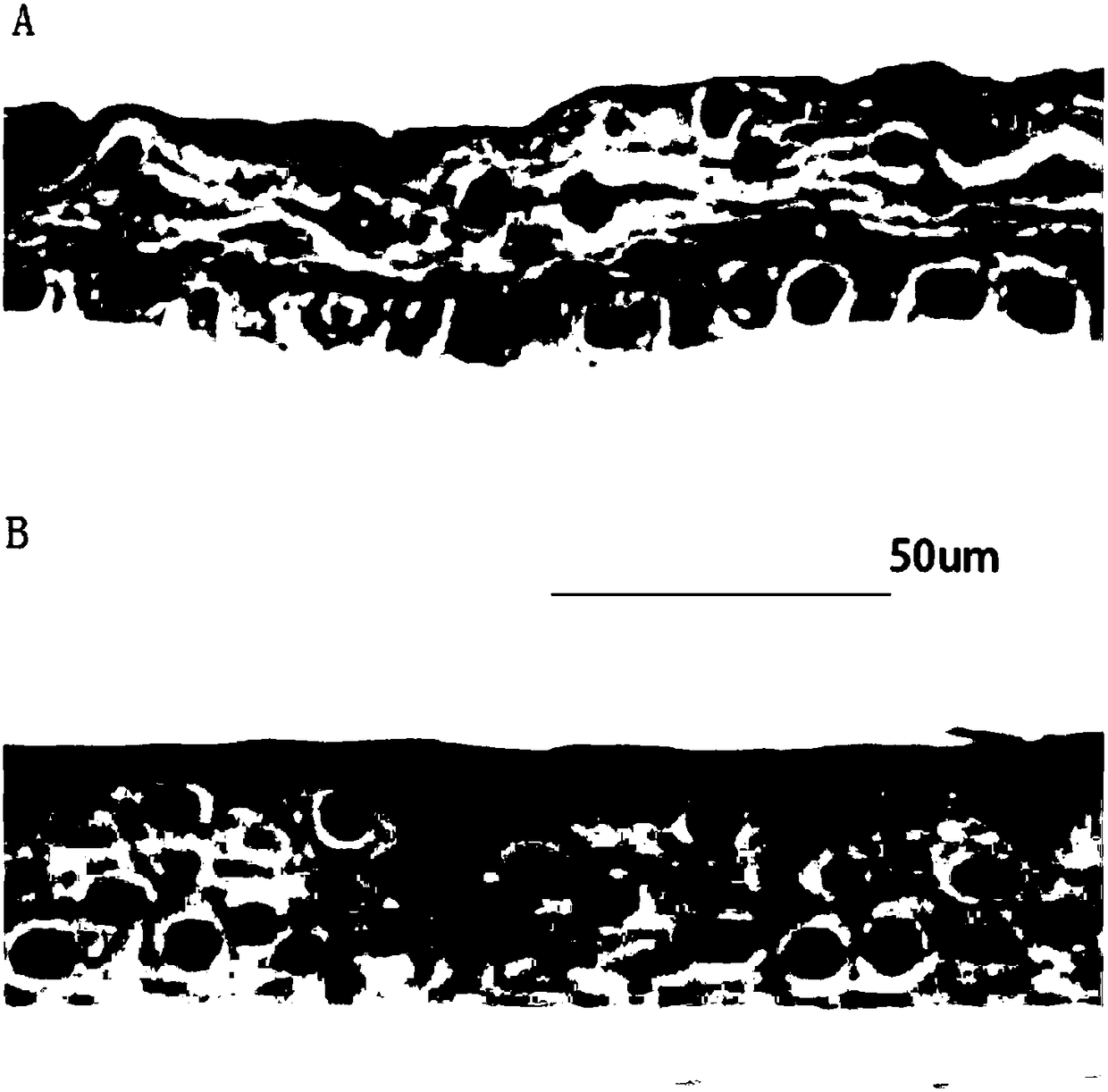
[0123] Culture of corneal endothelial cells

[0124] The culture seed cells of corneal endothelial cells are obtained from the corneas of other donors (the younger the better), and they are used within 10 days after donation.

[0125] The cornea is washed 2-5 times in phosphate-buffered saline containing 100U / ml penicillin and 100μg / ml streptomycin, 10-18min each time, after removing blood stains, carefully peel off the elastic membrane with the help of a vacuum suction device (Descemet membrane) and endothelial layer, the elastic membrane and endothelial layer after peeling will automatically roll up along the endothelial layer.

[0126] The collected posterior elastic membrane and endothelial layer were treated with collagenase (1mg / ml). After 2 hours, the corneal endothelial layer was partially separated. The complete separation of corneal endothelial cells requires collagenase treatment for approximately 6 hours (the time varies greatly depending on the donor), and the endotheli...


PUM


Abstract

The invention relates to the field of cell culture and particularly relates to a whole eye cornea bioengineering cultivation method. The various layers of the cornea are respectively cultivated, and the advantage is that only a cornea layer needed by a patient is cultivated when the patient does not need the transplantation of the whole cornea. According to the provided preparation method of the whole cornea, a seed cell between the cornea epidermal layer and a mesenchymal layer is an autologous cell, a corneal endothelial cell is from a donator, a large quantity of endothelial cells are obtained through cultivation, and the limit of the source of the donator is avoided. A collagen culture medium is taken as a gluing medium between the epidermal layer and the mesenchymal layer, and the intraneural growth of the human bioengineering cornea is promoted. The provided whole cornea and partial cornea both have an excellent optical property, the patients all recovers the eyesight after the operation, the cornea has a stable biochemical characteristic, the dissolution of the cornea in the later period does not exist, the biocompatibility is excellent, and no rejection reaction is caused.

Description

Technical field [0001] The present invention relates to the field of cell culture, in particular to a method for cultivating a full-thickness cornea of ​​the eye by bioengineering. Background technique [0002] Corneal disease has a high incidence rate in the world. Due to limited treatment methods, it is one of the main causes of blindness. At present, the main method of treating corneal diseases is full corneal or lamellar corneal transplantation. Due to the lack of qualified corneal donor sources, most patients who are blind or partially lost vision due to corneal diseases cannot receive effective treatment. [0003] In the past ten years, the development of biological tissue engineering technology has provided technical possibilities for in vitro construction of biological corneas. The bioreactor better simulates the living environment in the human body, and the use of seed cells and scaffold materials can prepare tissue engineered corneas in vitro; at the same time; Many tec...

Claims


Application Information

Patent Timeline
IPC IPC(8): C12N5/079
CPCC12N5/0621C12N2513/00C12N2533/30C12N2533/52C12N2533/54C12N2533/92
Inventor 张辉王旭左岩霞张兆蕾
Owner 山东麦德克斯生物科技有限公司
Why Eureka
Try Eureka