
3D bioprinting forming method

A bioprinting and molding technology, applied in tissue regeneration, medical science, prosthesis, etc., can solve the problems of material deformation, long preparation time, inability to meet scientific research and clinical needs, etc., achieve high speed, improve mechanical strength and The effect of chemical stability

Active Publication Date: 2018-08-28
THE FIRST AFFILIATED HOSPITAL OF SUN YAT SEN UNIV +1
View PDF6 Cites 3 Cited by
  • Summary
  • Abstract
  • Description
  • Claims
  • Application Information

AI Technical Summary

Problems solved by technology

Therefore, the final results of these methods are often unsatisfactory
[0005] Finally, limited by the physical properties of traditional soft tissue 3D printing inks (it is difficult to quickly process them into pre-designed shapes and structures), the existing soft tissue 3D printing technologies generally use cross-linking while printing, or first cross-linking and then The main disadvantages of the printing method include: ① need to use photo-crosslinkable materials, which limits the available materials, has biological safety hazards, and due to beam scattering, more serious problems will occur when preparing materials with smaller sizes. deformation; ②The crosslinking reaction takes a certain amount of time, making the structure of the material unstable during the printing process, and the overall preparation time is too long
[0006] In summary, the biological 3D soft tissue printing technology in the prior art is still immature and cannot meet the needs of scientific research and clinical practice.

Method used


Image

  • 3D bioprinting forming method
  • 3D bioprinting forming method

Examples

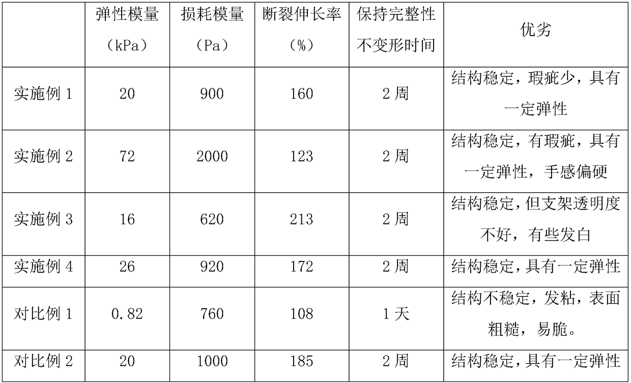
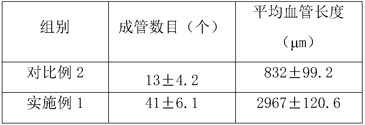

Embodiment 1

[0033] A kind of 3D printing biological ink, comprises the raw material of following mass percentage and is made of:

[0034] Extracellular Vesicle Suspension 10%

[0035] Pig dermal soft tissue derived extracellular matrix nanofiber powder 5%

[0036] Hydrochloric acid 1%

[0037] Ultrapure water balance.

[0038] The 3D printing bio-ink is prepared according to the following method:

[0039] Mix pig dermal soft tissue-derived extracellular matrix nanofiber micropowder, hydrochloric acid and ultrapure water, stir until completely dissolved, adjust pH to 6.5 with sodium bicarbonate, add extracellular vesicle suspension, mix evenly, and centrifuge to obtain Gel, the bioink material.

[0040] 3D printing forming method, comprising the steps of:

[0041] (1) 3D bioprinting: use the 3D bioprinter to print the composite ink material from the ink injection cylinder at 15°C through the needle to the receiving platform at 8°C according to the established digital model, and store ...

Embodiment 2

[0044] A kind of 3D printing biological ink, comprises the raw material of following mass percentage and is made of:

[0045] Extracellular vesicle suspension 20%;

[0046] Bovine dermis soft tissue-derived extracellular matrix nanofiber powder 15%;

[0047] Phosphoric acid 0.1%;

[0048] Ultrapure water balance.

[0049] The 3D printing bio-ink is prepared according to the following method:

[0050] Mix bovine dermis soft tissue-derived extracellular matrix nanofiber micropowder, phosphoric acid and ultrapure water, stir until completely dissolved, adjust pH to 6 with sodium phosphate, add extracellular vesicle suspension, mix well, and centrifuge to obtain coagulated Glue, the bio-ink material.

[0051] 3D printing forming method, comprising the steps of:

[0052] Carry out 3D printing according to the 3D printing molding method of Example 1, then wash with water, 0.5% (according to the amount of 3D printing bio-ink) glutaraldehyde cross-linking to set the shape.

Embodiment 3

[0054] A kind of 3D printing biological ink, comprises the raw material of following mass percentage and is made of:

[0055] Extracellular vesicle suspension 10%;

[0056] Pig dermis soft tissue-derived extracellular matrix nanofiber powder 8%;

[0057] Hydrochloric acid 1%;

[0058] Distilled water balance.

[0059] The 3D printing bio-ink is prepared according to the following method:

[0060] Mix pig dermal soft tissue-derived extracellular matrix nanofiber micropowder, hydrochloric acid and distilled water, stir until completely dissolved, adjust the pH to 7.45 with sodium hydroxide, add extracellular vesicle suspension, mix well, and centrifuge to obtain a gel , the bioink material.

[0061] 3D printing forming method, comprising the steps of:

[0062] (1) 3D bioprinting: use the 3D bioprinter to print the composite ink material from the ink injection cylinder at 20°C through the needle to the receiving platform at 4°C according to the established digital model, and...


PUM

PropertyMeasurementUnit
The average particle sizeaaaaaaaaaa

Abstract

The invention discloses a 3D bioprinting forming method. The 3D bioprinting forming method comprises the following steps: (1), performing 3D bioprinting: printing 3D printing bio-ink according to an established model by using a 3D bioprinter, and storing a printing bracket at low temperature to obtain a shaped gel material; (2), crosslinking: washing the shaped gel material by using water; then adding a crosslinking agent, and performing a crosslinking reaction under a low-temperature condition; finally, washing away the residual crosslinking agent by using water. By the 3D bioprinting formingmethod, low-temperature treatment and a crosslinking method are adopted for printing a target structure, so that rapid progress of 3D printing can be guaranteed, covalent bonds are further introducedinside collagen molecules and between the molecules, most of a collagen structure is retained, and the mechanical strength and the chemical stability of the printing material are improved, the absorption speed thereof in a body are controlled, and thus a collagen biomaterial plays a role conveniently.

Description

technical field [0001] The invention relates to the field of biotechnology, in particular to a 3D bioprinting molding method. Background technique [0002] The cases of 3D printing technology used in clinical medicine are no longer uncommon. The combination of this technology and medical treatment mainly includes the following types: ①preoperative diagnosis and surgical rehearsal model; ②implant preparation (bones, teeth, soft tissue organs, etc.) ; ③ External medical consumables (maxillofacial shaping braces, etc.). At present, the printing technology of hard tissue scaffolds such as bones and teeth is relatively mature, but the printing of soft tissue scaffolds still needs to be further improved. [0003] Most of the existing bio-inks available for soft tissue printing are polymer hydrogels, including artificially synthesized polymer materials and natural hydrogels such as collagen. The effect is weak; cells seeded on the printed scaffold only grow in situ throughout the...

Claims


Application Information

Patent Timeline
IPC IPC(8): A61L27/36A61L27/58B33Y10/00
CPCA61L27/3604A61L27/3633A61L27/58A61L2430/34B33Y10/00
Inventor 陈蕾李志勇祁少海陈永明吴军刘利新傅炀
Owner THE FIRST AFFILIATED HOSPITAL OF SUN YAT SEN UNIV
Learn More