Encoding gene of green fluorescent protein nano antibody and preparation method and application of encoding gene
A green fluorescent protein and nanobody technology, applied in the field of genetic engineering, can solve the problems of high production cost, poor stability, affecting the application of GFP antibody, etc., and achieve the effect of easy penetration of cell tissue, high specificity and affinity
- Summary
- Abstract
- Description
- Claims
- Application Information
AI Technical Summary
Problems solved by technology
Method used
Image
Examples
Embodiment 1
[0077] Embodiment 1: Construction of GFP nanobody library
[0078] (1) Prokaryotic expression of purified GFP, dilute the concentration to 1mg / mL, mix 1mg GFP with an equal volume of adjuvant (purchased from GERBU) for each immunization, and immunize an alpaca once every two weeks for a total of 5 Second-rate.
[0079] (2) After the 5 times of immunization, 50 mL of alpaca peripheral blood was drawn, blood lymphocytes were separated by dextran-diatrizoate meglumine density gradient centrifugation, and total cellular RNA was extracted by Trizol method.
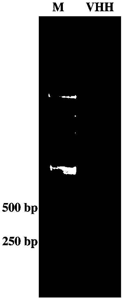
[0080] (3) According to TAKARA's PrimerScript TM 1st Strand cDNA Synthesis Kit manual, the extracted RNA is reverse-transcribed into cDNA, and the variable region (VHH region) of the alpaca heavy chain antibody is amplified by nested PCR.
[0081] The first round of PCR: perform 8 parallel PCRs, the primers used are as follows:
[0082] Upstream primer (5'-3'): GTCCTGGCTGCTCTTCTACAAGG,
[0083] Downstream primer (5'-3'):...
Embodiment 2
[0102] Example 2: Enrichment process of GFP-specific Nanobodies
[0103] (1) According to EZ-LinkTM Sulfo-NHS-LC-Biotinylation Kit (purchased from Thermo FisherScientific TM company), the purified GFP was coupled to biotin (biotin).
[0104] (2) Take 1 μg of biotin-coupled GFP and 30 μL of streptavidin-coupled magnetic beads (Dynabeads TM M-280 Streptavidin, Invitrogen) were incubated at room temperature for 30 min to bind GFP to the magnetic beads, and then washed 3 times with PBST to wash away unbound GFP.
[0105] (3) Add 500 μL phage display library (containing 5×10 12 A phage displaying the immune alpaca nanobody), and incubated at room temperature for 2h. Wash 25 times with PBST to wash away unbound or weakly binding phages. The phage specifically bound to GFP was dissociated with 500 μL trypsin (0.25 mg / mL), and 10 μL protease inhibitor cocktail (50x) (purchased from Roche Company) was added to the dissociated phage solution for neutralization.
[0106] (4) Take ...
Embodiment 3
[0111] Example 3: Enzyme-linked immunosorbent assay (ELISA) screening of GFP-specific nanobody positive monoclonal
[0112] (1) Randomly select 20 bacterial single clones from the overnight cultured Nanobody bacterial library obtained after the third round of screening in Example 2, inoculate them into LB medium respectively, cultivate to the logarithmic phase of bacterial growth, and add the final concentration 0.2mM IPTG, incubated overnight at 30°C to induce the expression of nanobodies.
[0113] (2) The bacteria were collected the next day, and CelLytic was used to TM B Cell Lysis Reagent (purchased from Sigma Company) was used to lyse the bacteria to obtain the nanobody crude extract. Take 100 μL of antibody crude extract, add it to the ELISA plate coated with GFP and blocked with 3% BSA, and incubate at room temperature for 1 hour.
[0114] (3) Wash 5 times with PBST, 1 min each time, to wash away unbound protein. Anti-pIII antibody (1:1000, purchased from NEB Compa...
PUM
 Abstract
Description
Claims
Application Information
 - R&D
- Intellectual Property
- Life Sciences
- Materials
- Tech Scout
- Unparalleled Data Quality
- Higher Quality Content
- 60% Fewer Hallucinations
Browse by: Latest US Patents, China's latest patents, Technical Efficacy Thesaurus, Application Domain, Technology Topic, Popular Technical Reports.
© 2025 PatSnap. All rights reserved.Legal|Privacy policy|Modern Slavery Act Transparency Statement|Sitemap|About US| Contact US: help@patsnap.com


