A microfluidic chip that captures rare blood cells
A microfluidic chip and chip technology, which can be used in pharmaceutical devices, enzymology/microbiology devices, bioreactors/fermenters for specific purposes, etc. Selective Offset, etc.
- Summary
- Abstract
- Description
- Claims
- Application Information
AI Technical Summary
Problems solved by technology
Method used
Image
Examples
Embodiment 1
[0086] Embodiment 1: the making of chip
[0087] 1. Mask production: Output the chip mask with a high-precision laser printer, and the size of the mask is 5-10×2-4cm.
[0088] 2. Production and exposure of photosensitive film: use photosensitive film with a thickness of 35 μm / sheet to build a convex skeleton for chip design graphics, routinely use 3-13 photosensitive films, stick the mask on the photosensitive film, and irradiate with ultraviolet light for 30 seconds- Expose for 120 seconds; use cleaning solution and a soft brush to carefully remove the dissolved photosensitive film after exposure, use an oscillator to shake evenly during cleaning to ensure that the remaining dissolved photosensitive film is fully removed, wash 3 times with RO water; then put Dry in an oven for 10-30 minutes to obtain the convex skeleton of the chip design pattern; observe the chip skeleton under a stereo microscope to ensure that the edge of the chip skeleton is smooth and the tiny spoiler is...
Embodiment 2
[0100] Embodiment 2: Detection of functional parameters of the chip
[0101]Create the grid model of the chip design in the Gambit2.4 version software, and use the ANSYS Fluent version 19.0 software to simulate the flow state of the liquid fluid in the chip. It can be seen from the simulation data that ① the fluid flow distribution in each flow channel is uniform, and the T-shaped deflector guides the liquid in the buffer pool to each flow channel more uniformly; The total flow velocity and the fluid density are set to "WATER-LIQUID", and the calculated flow velocity in each flow channel is 0.026-0.199m / s; ③ Karman vortex street is formed behind each spoiler column (such as Figure 5 and Figure 6 shown). Meet the design requirements.
Embodiment 3
[0102] Example 3: Detection of capture ability of breast cancer circulating tumor cells
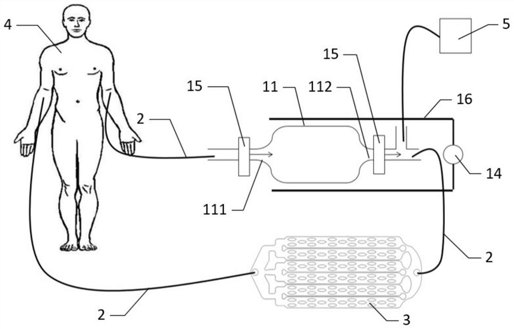
[0103] Insert the chip made in Example 1 into a container containing 10 ml of blood from a breast cancer patient. The blood enters the extracorporeal blood circulation system comprising the microfluidic chip set of the present invention after being guided out by blood vessels.
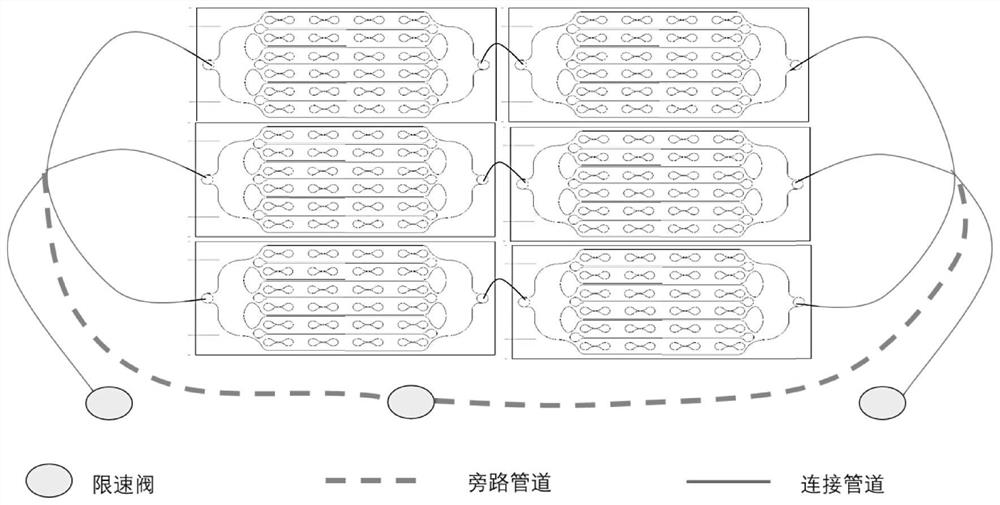
[0104] The microfluidic chipset composed of parallel / serial connection is connected through the medical-grade catheter, and the front-end anticoagulant slow-controlled release device is opened.
[0105] Turn on the power switch, and collect the cell suspension continuously for 1 hour.
[0106] Turn off the power switch of the device, turn off the automatic front-end anticoagulant slow-controlled release device, and the collection of circulating tumor cells ends.
[0107] Inject 200 μL of 1× PBS sterile solution to wash the chip 3 times.
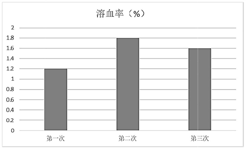
[0108] Observation of circulating tumor cells and hemolysis
[0109] Usi...
PUM
| Property | Measurement | Unit |
|---|---|---|
| diameter | aaaaa | aaaaa |
| diameter | aaaaa | aaaaa |
| diameter | aaaaa | aaaaa |
Abstract
Description
Claims
Application Information
 - R&D
- Intellectual Property
- Life Sciences
- Materials
- Tech Scout
- Unparalleled Data Quality
- Higher Quality Content
- 60% Fewer Hallucinations
Browse by: Latest US Patents, China's latest patents, Technical Efficacy Thesaurus, Application Domain, Technology Topic, Popular Technical Reports.
© 2025 PatSnap. All rights reserved.Legal|Privacy policy|Modern Slavery Act Transparency Statement|Sitemap|About US| Contact US: help@patsnap.com


