Two-photon fluorescent probe for detecting viscosity of positioning mitochondria and synthesizing method and application thereof

A fluorescent probe and synthesis method technology, applied in the field of organic small molecule fluorescent probes, can solve the problems of biological material damage, short excitation wavelength, etc., and achieve the effects of simple steps, convenient purification, and high yield

Inactive Publication Date: 2018-12-21
UNIV OF JINAN
View PDF1 Cites 15 Cited by
  • Summary
  • Abstract
  • Description
  • Claims
  • Application Information

AI Technical Summary

Problems solved by technology

[0004] Aiming at the problems in the prior art that the probes for detecting mitochondrial viscosity have short excitation wavelengths and are likely to cause damage to biol

Method used


Image

  • Two-photon fluorescent probe for detecting viscosity of positioning mitochondria and synthesizing method and application thereof
  • Two-photon fluorescent probe for detecting viscosity of positioning mitochondria and synthesizing method and application thereof
  • Two-photon fluorescent probe for detecting viscosity of positioning mitochondria and synthesizing method and application thereof

Examples


Embodiment 1

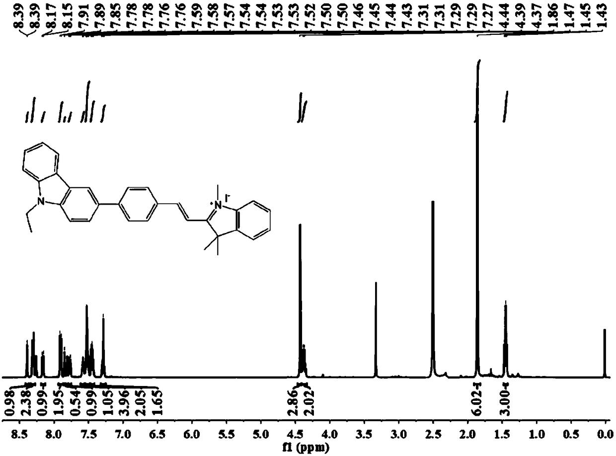
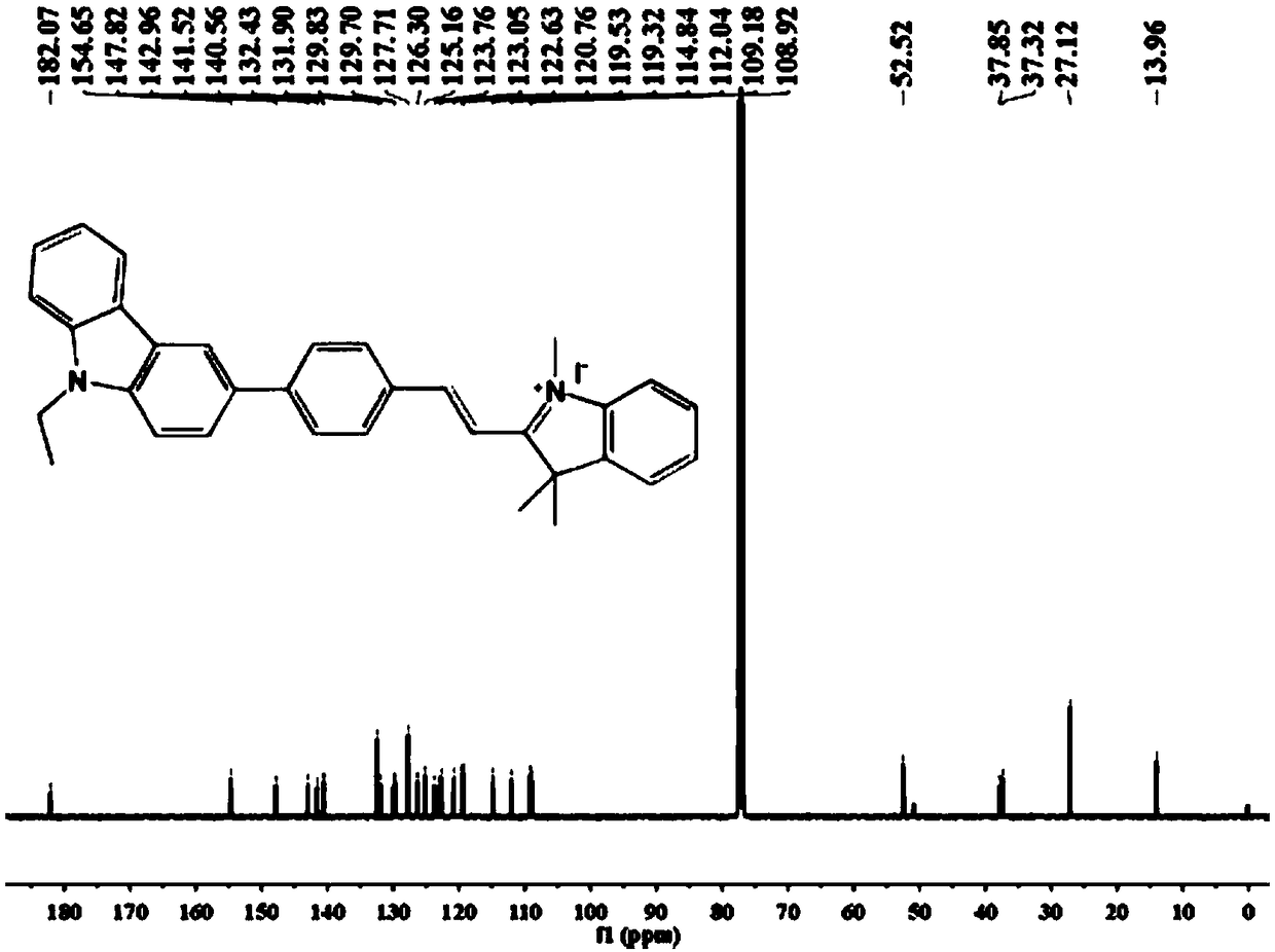
[0038] Example 1 Synthesis of Fluorescent Probes

[0039] (1) Synthesis of compound 3-(4-formylphenyl)-9-ethylcarbazole (3):

[0040] ;

[0041] Weigh 0.274 g 3-bromo-9-ethylcarbazole (1) (1 mmol), 0.18 g 4-formylphenylboronic acid (2) (1.2 mmol), 37 mg tetrakis-(triphenylphosphine)palladium ( 0.03 mmol), 0.445 g of potassium carbonate (3.23 mmol) was reacted in a mixed solvent of tetrahydrofuran and water = 3:1 (V:V), heated to 60 °C and refluxed, under N 2 After reacting for 12 h under atmospheric conditions, extract with dichloromethane, and remove a small amount of water remaining in the system with anhydrous sodium sulfate, then carry out vacuum distillation, spin dry the solvent to obtain a crude product, and use ethyl acetate with a volume ratio of 1:20 The ester and petroleum ether were used as the mobile phase for column chromatography separation and purification to obtain 3-(4-formylphenyl)-9-ethylcarbazole (3).

[0042] (2) Synthesis of compound iodide 1,2,3,3-...

Embodiment 2

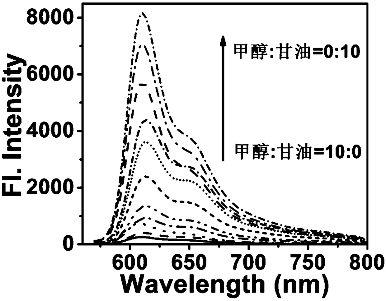
[0050] Example 2 Fluorescence spectra of fluorescent probe CBI-V in different viscosity systems

[0051] A test mother solution of dimethyl sulfoxide (DMSO) with a concentration of 1 mM fluorescent probe obtained in Example 1 was prepared for use.

[0052] In the test solution, take 3 ml of solvents with different ratios of glycerol and methanol (glycerol:methanol=0:10, 1:9, 2:8, 3:7, 4:6, 5:5, 6:4, 7: 3, 8:2, 9:1, 10:0), then add the probe mother solution (final concentration is 10 μM), carry out fluorescence scanning (excitation wavelength 520 nm, detection band 530-850 nm), measured in each system Relative fluorescence intensity, such as image 3 shown. Depend on image 3 It can be seen that with the increase of solvent viscosity, the relative fluorescence intensity becomes stronger.

Embodiment 3

[0053] Example 3 Colocalization imaging of fluorescent probes on mitochondria

[0054] A test mother solution of dimethyl sulfoxide (DMSO) with a concentration of 1 mM fluorescent probe obtained in Example 1 was prepared for use.

[0055] Seed an appropriate density of Hela cells into a sterilized 35 mm imaging dish in CO 2 Incubator (at 37°C, 5% CO 2 ), and after the cells adhered to the wall, add the nuclear dye DAPI, the viscosity fluorescent probe CBI-V and the commercial dye mitochondrial green to the cells at the same time, so that the final concentration of DAPI is 5 μM, and the final concentration of the viscosity fluorescent probe is 10 μM. μM, the final concentration of mitochondrial green is 5 μM. Half an hour later, discard the medium, wash the cells with PBS buffer three times, and then perform fluorescence imaging (excitation wavelength: 404 nm, blue channel: 425-475 nm; excitation wavelength: 488 nm, green channel: 500-550 nm; excitation Wavelength: 561 nm, r...


PUM


Abstract

The invention provides a two-photon fluorescent probe for detecting viscosity of positioning mitochondria and a synthesizing method and application thereof, and belongs to the field of small-moleculefluorescent probes. The structural formula of the two-photon viscosity probe is shown in the description; the probe can be prepared by reacting products of 3-bromine-9-ethyl carbazole and 4-formyl benzene boric acid and the products of 2, 3, 3-trimethyl-3H-benzpyrole and iodomethane. The probe has two-photon effect and is high in sensitivity relative to viscosity and is capable of detecting viscosity in a solution, cells and zebra fish body under a single-photon or a two-photon mode; and the probe has a wide application prospect in the field of biomolecule detection.

Description

technical field [0001] The invention belongs to the field of small organic molecule fluorescent probes, in particular to a fluorescent probe for detecting viscosity. Background technique [0002] Viscosity is the main factor to measure the fluidity and diffusibility of a thick fluid, and it is also the main reference index of the fluid diffusion rate. The viscosity of the microenvironment plays a very important role in pathological research, because the change of viscosity often affects the progress of various metabolisms in the cell microenvironment. Mitochondria are the major energy-producing regions in most eukaryotes, which play key roles in many important cellular processes such as ATP production, central metabolism and apoptosis. Viscosity changes in mitochondria may contribute to mitochondrial dysfunction. However, measuring viscosity at the microscopic scale, especially in tiny living cells, remains difficult. The local microviscosity in cells varies greatly from o...

Claims


Application Information

Patent Timeline
IPC IPC(8): C07D209/86G01N21/64A61K49/00
CPCA61K49/0032C07D209/86G01N21/6486
Inventor 林伟英阴军玲
Owner UNIV OF JINAN
Why Eureka
Try Eureka