Biological material, preparation method thereof and application of biological material as bone defect repairing material
A technology of biomaterials and bioceramic materials, applied in the application of bone defect repair materials, biomaterials and their preparation, can solve the problems of limited printing height, yellowing of materials, and low printing accuracy of biomaterials
- Summary
- Abstract
- Description
- Claims
- Application Information
AI Technical Summary
Problems solved by technology
Method used
Image
Examples
Embodiment 1
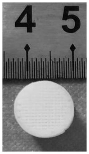
[0032] Example 1: C-CS 0.4g+Gel-MA 2.2g+HAP 5g+photocrosslinking+EDC / NHS crosslinking
[0033] The preferred embodiment provided by the present invention is an extruded 3D printing biomaterial for bone defect repair. The bio 3D printing paste formula includes an organic polymer part, an inorganic bioceramic particle part and a photoinitiating additive part.
[0034] The organic polymer part is composed of Gel-MA and C-CS. Take the C-CS 0.4g magnetic stirring dissolved in 11mLH 2 O, then add 2.2 g of the Gel-MA, and dissolve completely under stirring in a water bath at 50°C.
[0035] The photoinitiator additive part consists of photoinitiator 819 and photoinitiator 184 . Take the 0.15g of photoinitiator 819 and 0.25g of photoinitiator 184, and dissolve them in 0.4mL of acetone until they are completely dissolved, and the solution turns slightly yellow. The prepared photoinitiating additive is partially added to the organic polymer solution, and magnetically stirred until it ...
Embodiment 2
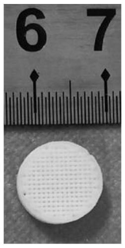
[0039] Example 2: C-CS 0.8g+Gel-MA 3.2g+photocrosslinking
[0040] In the biological 3D printing slurry component for bone repair of the present invention, the organic polymer part can be printed independently without relying on the inorganic particles.
[0041] Take 0.8g of the C-CS and dissolve it in 11mL of water with magnetic stirring, then add 3.2g of the Gel-MA, and dissolve completely in a 50°C water bath. The photoinitiator additive part consists of photoinitiator 819. Take 0.15 g of the photoinitiator 819 and dissolve it in 0.4 mL of acetone until completely dissolved.
[0042] The prepared photoinitiating additive is partially added to the organic polymer solution, and magnetically stirred until it dissolves completely to form a homogeneous solution.
[0043] Transfer the slurry to the barrel of the extrusion bioprinter, use a needle with an aperture of 0.16mm, set the air pressure to about 0.15MPa, the line speed to 6mm / s, set the line spacing to 0.3mm, and irradi...
Embodiment 3
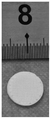
[0044] Example 3: C-CS 0.4g+Gel-MA 2.2g+HAP 3g+glutaraldehyde crosslinking
[0045] Take the C-CS 0.4g and dissolve it in 11mL H 2 O, then add 2.2 g of the Gel-MA, and dissolve completely in a water bath at 50°C.
[0046] The inorganic bioceramic particle part (hydroxyapatite) was sieved twice with a standard sieve of 0.15 mm aperture, 3 g of the sieved particles were taken, and the inorganic bioceramic particles were added to the above-mentioned homogeneous solution in five times, after each addition, Put it into a planetary centrifugal mixer and mix for about 15 minutes. After the last 3g is completely added, mix for 1 hour to obtain a uniform and stable extruded slurry.
[0047] Transfer the slurry to the barrel of the extrusion bioprinter, match the needle with the aperture of 0.41mm, set the air pressure to about 0.4MPa, the line speed to 8mm / s, the line spacing to 1.1mm, set the platform temperature to 4°C, and the barrel temperature to 30°C , to print. After printing...
PUM
 Abstract
Description
Claims
Application Information
 - R&D
- Intellectual Property
- Life Sciences
- Materials
- Tech Scout
- Unparalleled Data Quality
- Higher Quality Content
- 60% Fewer Hallucinations
Browse by: Latest US Patents, China's latest patents, Technical Efficacy Thesaurus, Application Domain, Technology Topic, Popular Technical Reports.
© 2025 PatSnap. All rights reserved.Legal|Privacy policy|Modern Slavery Act Transparency Statement|Sitemap|About US| Contact US: help@patsnap.com


