Fluorescent probe molecule ML-FP and preparation method and applications thereof

A fluorescent probe and molecular technology, applied in the field of fluorescent probes, can solve the problems of low toxicity and large Stokes shift, and achieve the effects of low cytotoxicity, large Stokes shift and high sensitivity

Active Publication Date: 2019-07-23
SOUTHEAST UNIV
View PDF3 Cites 6 Cited by
  • Summary
  • Abstract
  • Description
  • Claims
  • Application Information

AI Technical Summary

Problems solved by technology

Because it is a fluorophore in living organisms, green fluorescent protein has excellent biocompatibility, low toxicity, large Stokes shift, less interference from the bac...

Method used


Image

  • Fluorescent probe molecule ML-FP and preparation method and applications thereof
  • Fluorescent probe molecule ML-FP and preparation method and applications thereof
  • Fluorescent probe molecule ML-FP and preparation method and applications thereof

Examples


Embodiment 1

[0020] Example 1 Preparation and Characterization of Green Fluorescent Protein Fluorescent Probe Molecule ML-FP

[0021] (1) Preparation method of green fluorescent protein fluorescent probe molecule ML-FP

[0022] In an ice-water bath, 18 μL of triethylamine was slowly added dropwise to an anhydrous solution dissolved with morpholine indole fluorescent protein derivative M (47 mg, 0.084 mmol) and 2,4-dinitrobenzenesulfonyl chloride (27 mg, 0.101 mmol). CH 2 Cl 2 After the dropwise addition, the solution was gradually raised to room temperature, stirred and reacted for 12 hours, and the crude product was purified by column chromatography to obtain the fluorescent probe molecule ML-FP with a yield of 63%.

[0023] The synthetic route of compound ML-FP is as follows:

[0024]

[0025] Wherein the morpholine indole fluorescent protein derivative M was prepared according to literature method (Sarah Diab, AhmadM.Abdelaziz, Peng Li, Theodosia Teo, Sunita K.C.Basnet, Ben Noll, ...

Embodiment 2

[0032] Example 2 Application of green fluorescent protein fluorescent probe molecule ML-FP

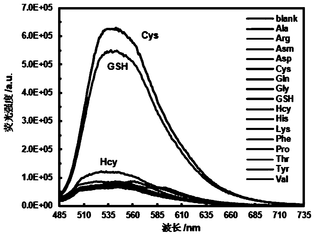
[0033]The maximum absorption wavelength of the probe molecule ML-FP is 470nm, the maximum emission wavelength is 542nm, and the Stokes shift is 72nm. The large Stokes shift makes the probe ML-GFP have excellent properties such as anti-background interference and high sensitivity. . At 25°C, incubate the probe ML-GFP with 5 times the equivalent of various biothiols and amino acids for more than 30 minutes, such as L-alanine (Ala), L-arginine (Arg), L-day Partine (Asm), L-Aspartic Acid (Asp), Cysteine ​​(Cys), L-Glutamine (Gln), Glycine (Gly), Glutathione (GSH), Homocysteine Acid (Hcy), L-Histidine (His), L-Lysine (Lys), L-Phenylalanine (Phe), Proline (Pro), L-Threonine (Thr), Tyrosine amino acid (Tyr) and valine (Val). figure 1 In the buffer solution of the fluorescent probe molecule ML-GFP (the pH of the buffer solution is 7.2, the solvent is a mixed solvent composed of water and et...

Embodiment 3

[0034] Example 3 Application of lysosome targeting active thiol fluorescent probe of green fluorescent protein fluorescent probe molecule ML-FP

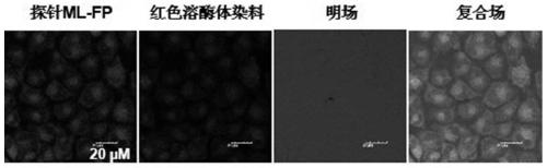
[0035] The probe molecule ML-FP has good cell permeability, can be quickly located in lysosomes, and can quantitatively monitor biothiols in lysosomes. figure 2 Lysosome co-localization of probe molecule ML-FP and commercial red lysosome localization reagent Lyso-Tracker Red (Nanjing Lattice Chemical Reagent Company) in Bel-7402 cells (purchased from Nanjing Lattice Chemical Reagent Company) Imaging photos. The red lysosome localization reagent Lyso-Tracker Red and ML-FP were simultaneously injected into Bel-7402 living cells for co-staining, and the cells were incubated in an incubator for 30 minutes. After washing the cells three times with PBS buffer solution, the fluorescent signals under the green field and red field were observed by confocal microscope. Such as figure 2 As shown, cells with green and red fluorescent signal...


PUM


Abstract

A fluorescent probe molecule ML-FP disclosed by the invention comprises a green fluorescent protein-sulfonate structure and a morpholine lysosome orientating group. The invention also discloses a preparation method and applications of the fluorescent probe molecule ML-FP. The fluorescent molecule can be used as a lysosome orientating active mercaptan fluorescent probe. The fluorescent probe is a fluorescent molecule prepared by reacting a morpholine-indole fluorescent protein derivative with 2,4-dinitrobenzenesulfonyl chloride. An aryl sulfonate structure is removed by an aromatic nucleophilicsubstitution reaction selectively induced by biological mercaptan so as to switch a fluorescence switch to realize selective recognition of cysteine and glutathione. The fluorescent probe molecule has the advantages of strong anti-background interference ability, high sensitivity, low cytotoxicity and good biocompatibility, can quickly enter lysosome, can be used as the lysosome orientating active mercaptan fluorescent probe, and has wide applications in fluorescence imaging, bioluminescence analysis, medicine research and development, fluorescence labeling and endogenous or exogenous activemercaptan detection.

Description

technical field [0001] The invention relates to the technical field of fluorescent probes, in particular to a fluorescent probe molecule ML-FP and its preparation method and application, in particular to a lysosome-localized green fluorescent protein active thiol fluorescent probe and its preparation method and application. Background technique [0002] Biothiol small molecules such as glutathione (GSH), cysteine ​​(Cys), and homocysteine ​​(GSH) play an important role in living organisms, participating in many physiological balance and metabolic processes of the human body, maintaining normal physiological functions of the human body. The content level changes of these small active thiol molecules in the body are closely related to physiological health. For example, cysteine ​​(Cys), which is not only a precursor of glutathione, acetyl coenzyme and taurine, but also a provider of sulfur ligands in the sulfur-iron complex in the body, can also weaken the body's absorption o...

Claims


Application Information

Patent Timeline
IPC IPC(8): C07D403/06G01N21/64
CPCC07D403/06G01N21/6428G01N21/643
Inventor 钱鹰沈宝星支旭朱雯瑜
Owner SOUTHEAST UNIV
Why Eureka
Try Eureka