
A core-shell structure microsphere and its application for monitoring the mechanical properties and contraction frequency of muscle cells

A technology with mechanical properties and core-shell structure, applied in the field of biomedicine, it can solve the problems of not being able to fully simulate muscle cells, accurately reproducing the connections of cells in three-dimensional tissues and the microenvironmental obstacles in which they are located, and achieving good biocompatibility, High detection sensitivity and wide range of materials

Active Publication Date: 2020-11-10
ZHEJIANG UNIV
View PDF2 Cites 0 Cited by
  • Summary
  • Abstract
  • Description
  • Claims
  • Application Information

AI Technical Summary

Problems solved by technology

However, the growth of cells is regulated by many factors. Traditional 2D cell culture cannot completely simulate the in vivo environment of muscle cells, and there are some obstacles in accurately reproducing the connection of cells in three-dimensional tissues and the microenvironment they are in.

Method used


Image

  • A core-shell structure microsphere and its application for monitoring the mechanical properties and contraction frequency of muscle cells
  • A core-shell structure microsphere and its application for monitoring the mechanical properties and contraction frequency of muscle cells
  • A core-shell structure microsphere and its application for monitoring the mechanical properties and contraction frequency of muscle cells

Examples


Embodiment 1

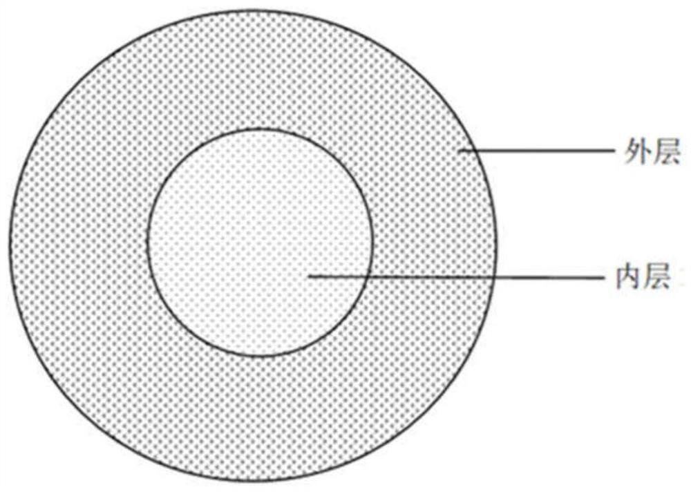
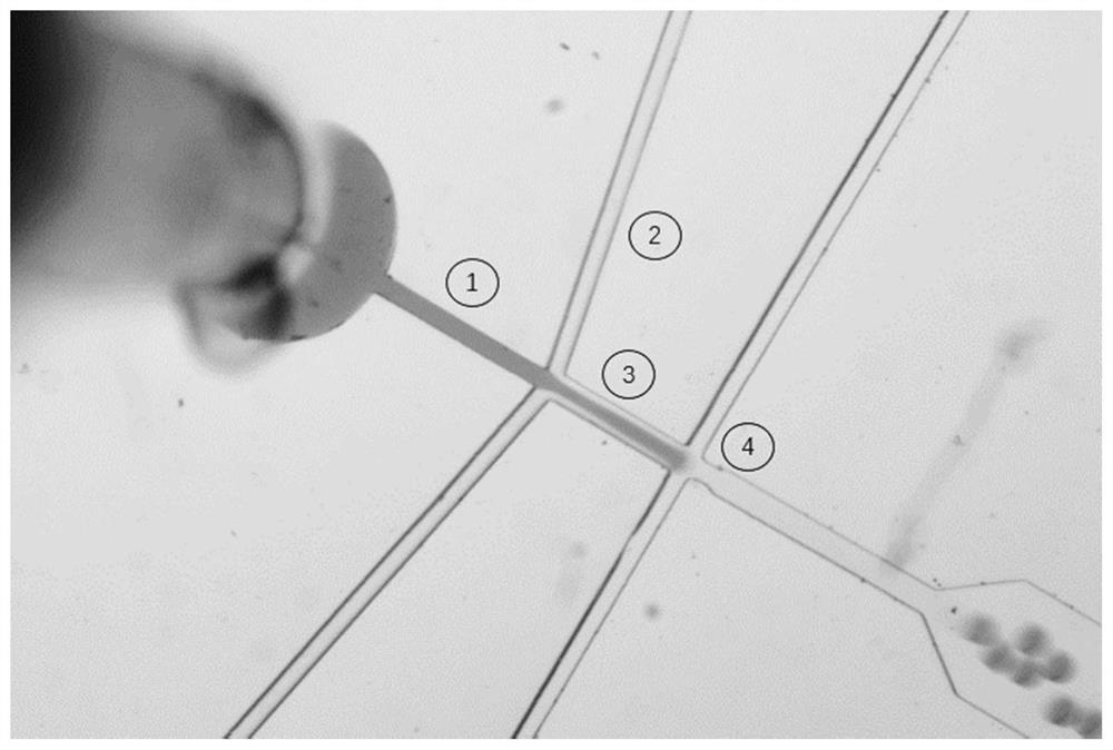
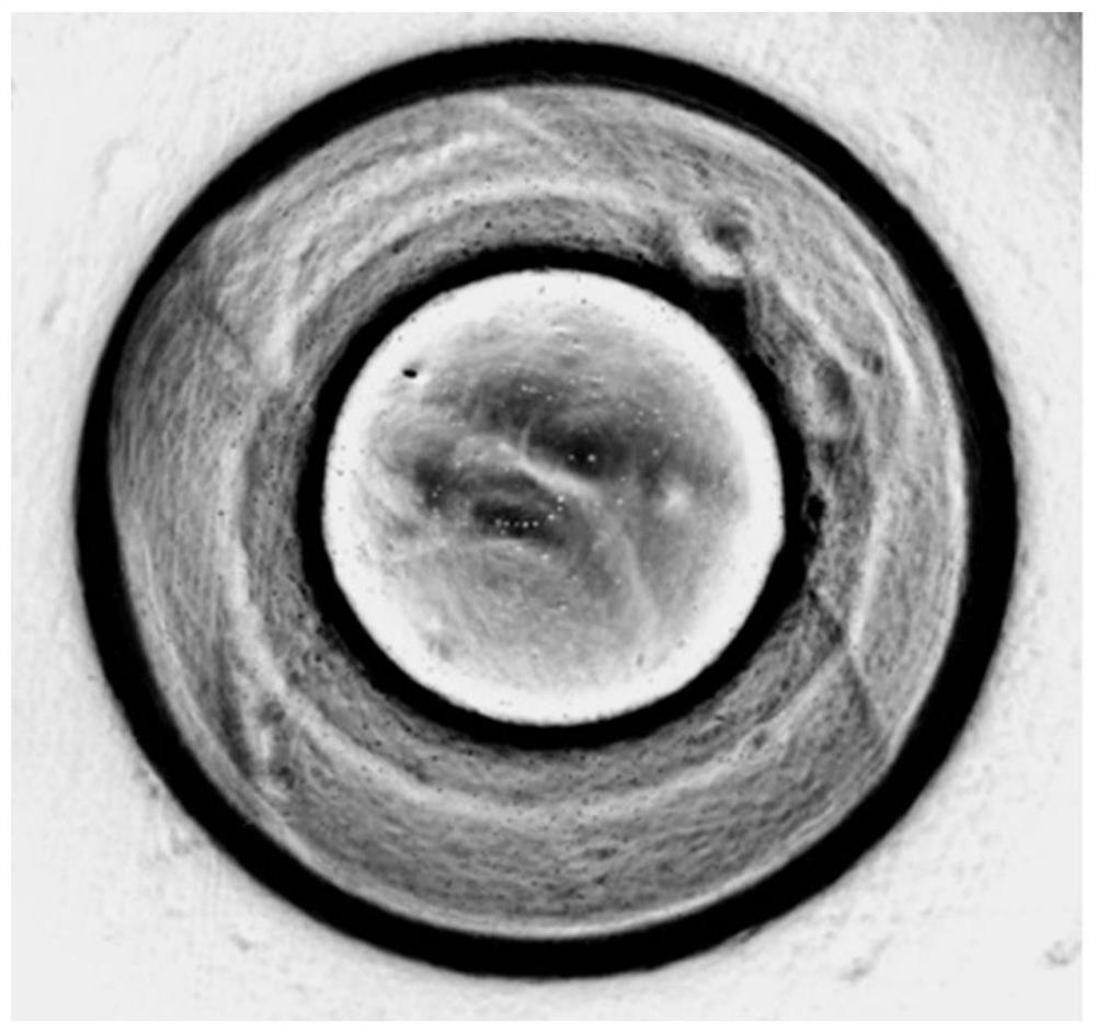
[0027] Using a microfluidic chip to prepare a figure 1 Microspheres with a core-shell structure are shown. First prepare the core and shell materials, the core material is 3% FITC-labeled agarose gel solution; the shell material is the cardiomyocytes obtained by induction and differentiation of human pluripotent stem cells (10 5 / mL) and Gel-MA gel solution (4%, w / w) mixture. Use a syringe pump to feed the core and shell materials into the figure 2 In the channels 1 and 2 of the microfluidic chip shown, the two materials form an interlayer flow at 3, and are pinched off by the oil phase at 4 to form droplets. The liquid flow rates at channels 1 and 2 are 20 μl / hour, 60 microliters / hour, and the oil phase velocity at channel 4 is 180 microliters / hour. Collect the droplets and incubate them in a 37°C incubator for 30 minutes. After the gel of the shell and core layers solidifies, microspheres with a core-shell structure are formed. The oil phase is discarded and the microsp...

Embodiment 2

[0030] Microspheres with a core-shell structure were prepared using a coaxial nozzle. Preparation of core and shell materials, core material: 80% (w / w) benzyl silicone oil, 12% vinyl silicone oil, 6% hydrogen-containing silicone oil, 1% platinum catalyst, 1% CdSe quantum dots; shell material: Cardiomyocytes induced and differentiated from human pluripotent stem cells (10 6per mL), gelatin solution (4%, w / w), transglutaminase (5 mg / mL). The core and shell materials are respectively extruded from the inner and outer layers of the coaxial nozzle, and the flow rates of the inner and outer layers are 40 microliters / hour and 20 microliters / hour, respectively. Apply a high voltage of 3.5KV to separate the droplets from the nozzle and fall into the high-viscosity silicone oil below (viscosity 2000cst). Put it in a 37°C incubator and incubate for 30 minutes. After the shell gel is solidified, discard the silicone oil, add the medium, centrifuge to separate the microspheres, add them ...

Embodiment 3

[0035] Using a microfluidic chip to prepare a figure 1 Microspheres with a core-shell structure are shown. First prepare the core and shell materials, the core material is 3% FITC-labeled agarose gel solution; the shell material is the cardiomyocytes obtained by induction and differentiation of human pluripotent stem cells (10 7 individual / mL) and gelatin solution (4%, w / w), the mixture of glutaminase (5mg / mL). Use a syringe pump to feed the core and shell materials into the figure 2 In the channels 1 and 2 of the microfluidic chip shown, the two materials form a sandwich flow at 3, and are pinched off by the oil phase at 4 to form droplets. The liquid flow rates at channels 1 and 2 are 10 μl / hour, 50 microliters / hour, and the oil phase velocity at channel 4 is 200 microliters / hour. Collect the droplets and incubate them in a 37°C incubator for 30 minutes. After the gel of the shell and core layers solidifies, microspheres with a core-shell structure are formed. The oil p...


PUM

PropertyMeasurementUnit
diameteraaaaaaaaaa
thicknessaaaaaaaaaa
particle diameteraaaaaaaaaa

Abstract

The invention discloses core-shell-structured microspheres for monitoring mechanical properties and contraction frequency of myocytes as well as application thereof, and belongs to the technical fieldof biomedicine. At present, studies on cell contraction force are mainly based on two-dimensional cell models, so that in vivo environment of myocytes cannot be completely simulated. The invention provides three-dimensional microspheres with core-shell structure. Each of the three-dimensional microspheres with core-shell structure comprises a core layer and a shell layer; the core layer is a sphere formed by performing solidification on a first gel material; the shell layer is a shell composed of cells and a second gel material, and wraps the core layer; and the core layer deforms along withpulsation of the cells in the shell layer, so that, distribution of forces produced by the shell-layer cells on the core layer can be obtained by observing the deformation of the core layer. Comparedwith two-dimensional cell culture, the three-dimensional cell culture system focuses on cell-to-cell contact and cell-to-matrix contact, so that, growth environment of organisms can be better simulated; and thus, the three-dimensional microspheres with core-shell structure can be applied in disease research cell models and drug screening models.

Description

technical field [0001] The invention relates to the field of biomedicine, in particular to a microsphere with a core-shell structure for monitoring the mechanical properties and contraction frequency of muscle cells and its application. Background technique [0002] Cells are the basic units that make up the structure and function of organisms, the basic unit of life activities, and the smallest entities that show all the characteristics of life states. Any life activities of the human body are closely related to cells. Cell mechanical properties are closely related to cell life activities, tissues, organs, and physiological processes of the body. Therefore, by quantifying the changes in the mechanical properties of cells, it can help humans better understand the life processes and regulatory mechanisms of cells, and can also play a role in drug screening and disease diagnosis. [0003] In recent years, the measurement of cell mechanical properties accompanied by muscle cel...

Claims


Application Information

Patent Timeline
Patent Type & Authority Patents(China)
IPC IPC(8): C12N11/10C12N11/04C12Q1/02B01J13/22
CPCB01J13/22C12N9/1044C12N11/04C12N11/10C12N2503/02C12Y203/02013G01N33/5008G01N33/5064G01N2500/10
Inventor 王平贺川江左一鸣贾怡萱赵梦雨刘梦雪
Owner ZHEJIANG UNIV
Learn More