In-vitro liver function detection method and system

A liver function and detection method technology, which is applied in the field of medical diagnosis, can solve the problems that static values ​​cannot be used to reflect liver function, large individual differences in indicators, and large deviations in physiological indicators, etc., and achieve the effect of overcoming the inability to quantitatively detect liver function

Active Publication Date: 2020-06-09
GENERAL HOSPITAL OF SOUTHERN THEATRE COMMAND OF PLA +2
View PDF7 Cites 1 Cited by
  • Summary
  • Abstract
  • Description
  • Claims
  • Application Information

AI Technical Summary

Problems solved by technology

However, the current application of these contrast agents is limited to improving the imaging contrast between liver tissue and tumor tissue during MRI imaging, and is mostly used to detect small solid tumors in the liver.
[0005] The following obstacles still exist in the liver function test of isolated liver donors: (1) Most of the in vivo liver function test indicators cannot directly reflect the metabolic function of the isolated liver. No direct relationship; (2) The indicators in the in vitro detection of perfusate have large individual differences, and the deviation from normal physiological indicators is large, so it is difficult to quantitatively evaluate the isolat

Method used


Image

  • In-vitro liver function detection method and system
  • In-vitro liver function detection method and system
  • In-vitro liver function detection method and system

Examples


Embodiment 1

[0054] In the experiment with 35-45kg Bama pigs, blood-donating pigs (n=6) were anesthetized and placed in the abdominal aorta and inferior vena cava, and about 1500ml of blood was released. The blood was filtered for white blood cells, and then stored at 4°C. Donor liver pigs (n=6) were anesthetized and placed catheters in the abdominal aorta, inferior vena cava, and portal vein, and instilled UW solution at 4°C through the abdominal aorta and portal vein. The dissociated liver was taken out and stored in UW solution at 4°C. Then the arteries, veins, and bile ducts of the liver are connected to the relevant pipelines of the isolated liver mechanical perfusion apparatus, and the pipelines are pre-filled with perfusate. The composition of the perfusion fluid is: 1.5L of whole blood, 150ml of 4% serum albumin, 18ml of 3% sodium bicarbonate, 9ml of 8% calcium chloride, 4000U heparin, 1g of cefoxitin, 500mg of metronidazole, and 15mg of bile salts; In addition, according to the p...

Embodiment 2

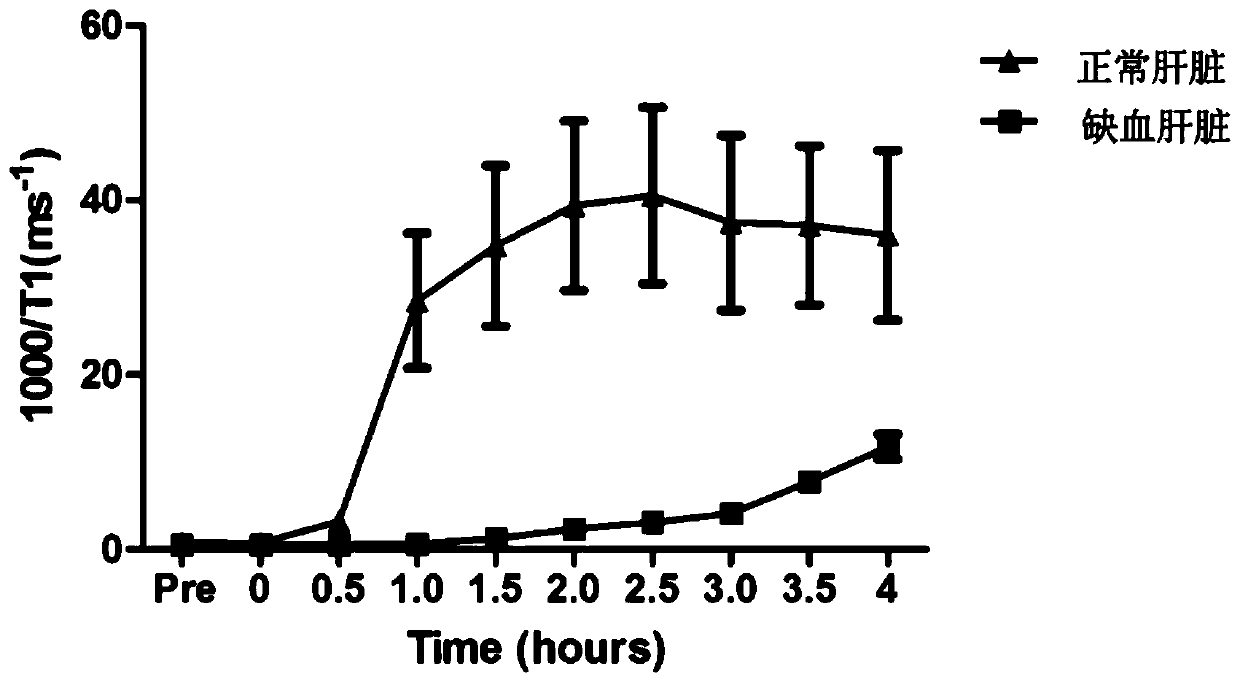
[0058] Normal group: 35-45kg Bama pigs were used for the experiment. The blood-donating pigs (n=6) were anesthetized and placed in the abdominal aorta and inferior vena cava, and about 1500ml of blood was released. The blood was filtered for white blood cells, and then stored at 4°C. Donor liver pigs (n=6) were anesthetized and placed catheters in the abdominal aorta, inferior vena cava, and portal vein, and instilled UW solution at 4°C through the abdominal aorta and portal vein. The dissociated liver was taken out and stored in UW solution at 4°C. Then the arteries, veins, and bile ducts of the liver are connected to the relevant pipelines of the mechanical perfusion apparatus for the isolated liver, and the pipelines are pre-filled with the above-mentioned perfusate. The perfusate consists of: 1.5L of whole blood, 100ml of 6% serum albumin, 24ml of 2% sodium bicarbonate, 5ml of 12% calcium chloride, 6000U of heparin, 1g of cefoxitin, 500mg of metronidazole, and 25mg of bile...

Embodiment 3

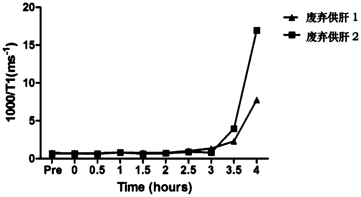
[0064] Abandoned human liver donors (n=2) were stored in cold storage for about 6 hours, taken out from the 4°C cold storage box, and the arteries, veins, and bile ducts of the liver were connected to the relevant pipelines of the isolated liver mechanical perfusion apparatus. Fill with the above perfusate. The composition of the perfusion solution is: 1.5L of whole blood, 130ml of 5% serum albumin, 21ml of 2.5% sodium bicarbonate, 7ml of 10% calcium chloride, 5000U heparin, 1g of cefoxitin, 500mg of metronidazole, and 20mg of bile salts; In addition, according to the pH of the perfusate, regular insulin and 2.5% sodium bicarbonate were used as appropriate, and the pH of the perfusate was adjusted within the range of 7.3 to 7.5.

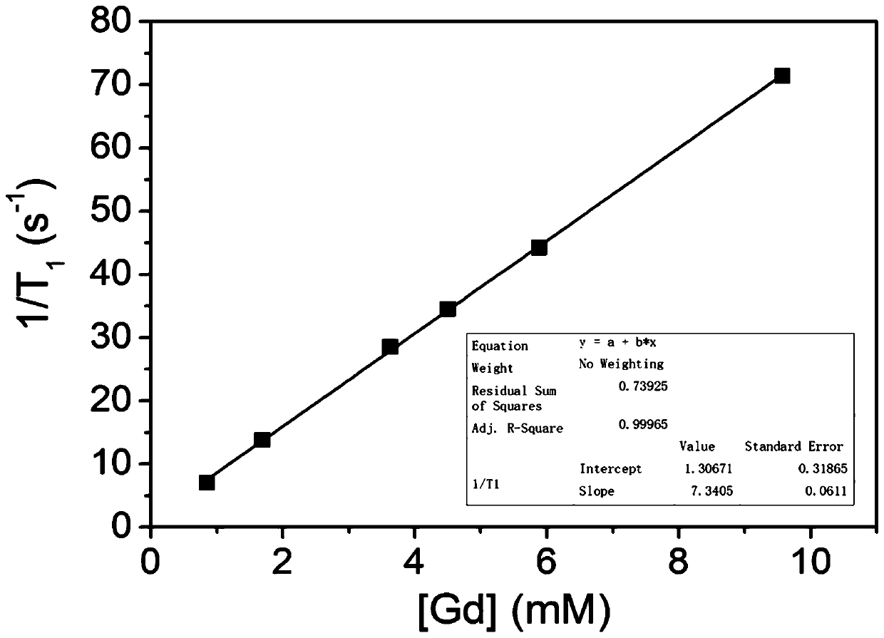
[0065] After 2 hours of perfusion, when the bile outflow is stable, inject gadoxetic acid disodium into the portal circulation, 0 hour, 0.5 hour, 1 hour, 1.5 hour, 2 hours, 2.5 hours, 3 hours, 3.5 hours, 4 hours, After 4.5 hours and 5 hours, take ou...


PUM

PropertyMeasurementUnit
Magnetic field strengthaaaaaaaaaa
Concentrationaaaaaaaaaa

Abstract

The invention relates to the technical field of medicine diagnosis and in particular relates to an in-vitro liver function detection method and system. The method comprises the following steps: a) respectively accessing arteries, veins and biliary tracts of a liver into related pipelines of an in-vitro liver mechanical perfusion apparatus, wherein the pipelines are filled with whole blood in advance; b) injecting a contrast medium into a hepatic portal vein to implement circulation, and testing T1 relaxation time of discharged bile by using a low-field magnetic resonance imaging analyzer; andc) comparing variation situations of the T1 relaxation time of the discharged bile with a reference value, and judging that if the T1 relaxation time of a same time point is longer than the referencevalue, it means that the metabolic function of the liver is lower that a liver corresponding to the reference value, and otherwise, it means the metabolic function of the liver is better than that ofthe liver corresponding to the reference value. The method makes up the limitation that liver functions cannot be directly reflected by conventional detection indexes.

Description

technical field [0001] The invention relates to the technical field of medical diagnosis, in particular to a method and system for detecting the function of an isolated liver. Background technique [0002] Liver transplantation is a gradually mature medical technology. The process includes: after the donor (donor) donates the liver, the liver is taken out and stored in a refrigerator and transported to the transplant medical center, and then the liver is surgically transplanted into the patient (recipient). Donor livers inevitably experience cold and warm ischemic injury during acquisition, storage, and transportation. A certain degree of cold and warm ischemic injury will lead to problems such as graft failure and biliary complications; other livers come from donors who are elderly or have fatty liver, which will seriously affect the postoperative survival and prognosis of transplanted patients. Quality assessment of donor livers prior to transplantation is critical. The...

Claims


Application Information

Patent Timeline
IPC IPC(8): A61B5/00A61B5/055
CPCA61B5/4244A61B5/055A61B5/4866
Inventor 欧阳青梁国海霍枫谭晓宇邝伟健
Owner GENERAL HOSPITAL OF SOUTHERN THEATRE COMMAND OF PLA
Why Eureka
Try Eureka