Zika virus E protein conjugate vaccine and preparation method thereof

A protein-binding vaccine, Zika virus technology, applied in the fields of botanical equipment and methods, biochemical equipment and methods, viruses, etc., can solve problems such as long-term toxicity, non-degradability, and limited clinical application of nanoparticle vaccines

Active Publication Date: 2020-08-11
INST OF PROCESS ENG CHINESE ACAD OF SCI
View PDF4 Cites 4 Cited by
  • Summary
  • Abstract
  • Description
  • Claims
  • Application Information

AI Technical Summary

Problems solved by technology

However, nanoparticle materials such as carbon nanotubes and gold nanoparticles have poor water solubility, are not easily degraded, have long-term toxicity, and are not easy to scale up, which limit the clinical application of nanoparticle vaccines.

Method used


Image

  • Zika virus E protein conjugate vaccine and preparation method thereof
  • Zika virus E protein conjugate vaccine and preparation method thereof
  • Zika virus E protein conjugate vaccine and preparation method thereof

Examples


Embodiment 1

[0074] Embodiment 1: Expression and purification of E protein

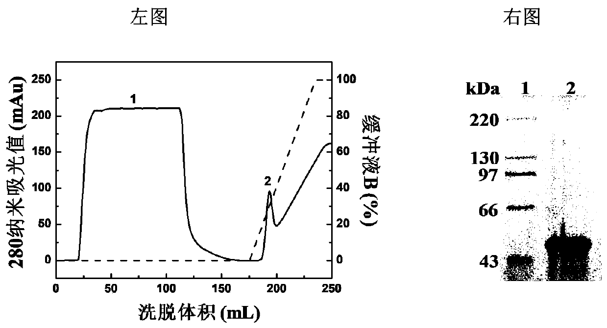
[0075] The gene sequence of the E protein is derived from the envelope protein of the Zika virus BeH818995 strain (Genebank number: KU365777); 6 histidine residues are fused to the C-terminus of the E protein gene sequence source (1-409 amino acid residues). After the synthetic gene sequence was cloned into pET21a expression vector, it was transformed into Escherichia coli BL21(DE3) for high-level expression. Synthetic DNA sequences were determined by restriction enzyme digestion and DNA sequence analysis. Activate Escherichia coli cells expressing E protein, inoculate 200ml LB medium at a ratio of 1%, and culture with shaking until OD 600 Equal to 0.6-0.8, add IPTG with a final concentration of 0.5mmol / L, and induce at 37°C for 3-4 hours. Centrifuge at 4° C. and 10000 rpm for 10 minutes, collect the bacterial cells, and resuspend in 50 mM Tris-HCl buffer (pH 8.0). Ultrasonication was performed for 1 hour in an...

Embodiment 2

[0078] Embodiment 2: Preparation and purification of vaccine

[0079] (1) Preparation of E-PS-1
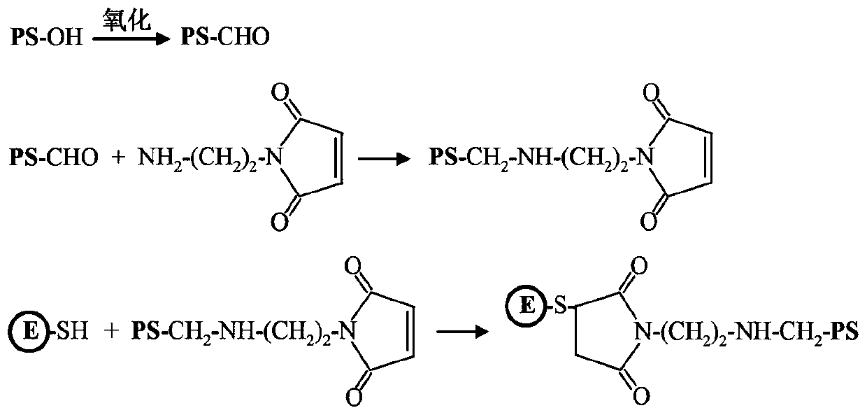
[0080] E protein has a cysteine ​​residue located on the surface of the E protein molecule; the sulfhydryl group of cysteine ​​can be used to covalently bind β-glucan. Such as figure 2 Sodium periodate was dissolved in a volume of 5 mL of 50 mM acetate buffer (pH 5.6) to a final concentration of 40 mM. β-glucan (PS-OH) was dissolved in a volume of 5 ml of 50 mM acetate buffer (pH 5.6) to make a final concentration of 10 mg / mL. The two were mixed and placed at room temperature in the dark for 20 minutes. Subsequently, unreacted sodium periodate was removed by dialysis against PBS buffer (pH 7.4). Oxidized β-glucan (5 mL) with a concentration of 2 mg / mL and aminoethyl-maleimide ester (5 mL) with a concentration of 0.2 mg / mL were reacted overnight in PBS buffer (pH 7.4), and the reaction temperature is 4°C. Unreacted aminoethyl-maleimide ester was removed by dialysis against P...

Embodiment 3

[0085] Embodiment 3: the purity of gel filtration analysis vaccine

[0086] E-PS-1 and E-PS-2 were identified by analytical Superdex 200 gel filtration column (1.0cm×30cm), the eluent was PBS buffer (pH 7.4), and the flow rate was 0.5mL / min. Such as Figure 5 As shown, E protein, E-PS-1 and E-PS-2 all present a single elution peak. Compared with the elution peak of E protein (15.4mL), the elution time of E-PS-1 and E-PS-2 was obviously earlier. This indicates that covalent attachment to β-glucan significantly enhances the hydration volume of E protein. The peak elution time of E-PS-1 and E-PS-2 was 7.7mL, indicating that the hydrazone bond and disulfide bond as connecting bridges did not change the hydration volume of the conjugate.


PUM


Abstract

The invention discloses a Zika virus E protein conjugate vaccine and a preparation method thereof. The preparation method comprises the following specific steps of: 1) oxidizing an ortho-position hydroxyl group of [beta]-glucan into an aldehyde group, so as to obtain oxidized [beta]-glucan; 2) adding a connecting bridge compound, enabling the connecting bridge compound to react with the aldehyde group in the oxidized [beta]-glucan to obtain [beta]-glucan containing a connecting bridge; and 3) enabling Zika virus E protein to react with the [beta]-glucan containing the connecting bridge, and carrying out purification to obtain the Zika virus E protein conjugate vaccine. The vaccine can be induced to generate a high-level cellullar immunologic response and a body fluid immunologic response.

Description

technical field [0001] The invention relates to the field of biomedicine, in particular, the invention relates to a Zika virus recombinant protein-conjugated vaccine based on β-glucan modification. Background technique [0002] Zika virus is an RNA virus with an icosahedral structure and an envelope, belonging to the flavivirus genus. Zika virus is transmitted by Aedes mosquitoes, can infect nerve cells, and break through the blood fetus, blood eye, blood testis and blood brain barrier, causing microcephaly in newborns and Guillain-Barré syndrome in adults. In recent years, Zika outbreaks in Central and South America and even around the world have shown unusually severe clinical manifestations. Due to the close personnel exchanges between my country and Zika virus endemic areas, the continuous occurrence of Zika virus imported cases poses a serious threat to my country's public health security. Globally, there is currently no effective means to prevent and control Zika vir...

Claims


Application Information

Patent Timeline
Patent Type & Authority Applications(China)
IPC IPC(8): A61K39/12A61K39/39A61K39/385A61K39/215A61P31/14C07K14/18C12N15/40C12N15/70
CPCA61K39/12A61K39/39A61K39/385A61P31/14C07K14/005C12N15/70A61K2039/55583A61K2039/575A61K2039/60C12N2770/24134C12N2770/20034C12N2770/24122Y02A50/30
Inventor 胡涛齐金铭于卫立申莉娟
Owner INST OF PROCESS ENG CHINESE ACAD OF SCI
Why Eureka
Try Eureka